Rehabilitation of Hand Burn Injuries
Top Contributors - Rania Nasr, Lucinda hampton, Shaimaa Eldib, Kim Jackson, Aya Alhindi, Nikhil Benhur Abburi, Anas Mohamed and Chelsea Mclene
Introduction[edit | edit source]
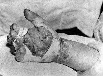
The importance of rehabilitation of burn injuries has been increased due to the improved short and long survival rate of people with large burns. Successful outcomes following hand burn injury require an understanding of the rehabilitation needs of the patient. Rehabilitation of hand burns begins on admission, and each patient requires a specific plan for range of motion and/or immobilization, functional activities, and modalities. The rehabilitation care plan typically evolves during the acute care period and during the months following injury[1].
- Burn injuries in hands are complex and the appearance of contractures is a common complication.
- Hand burn injuries often result in limited hand function especially flexion/extension of fingers and present a major hindrance in rehabilitation. These injuries also decline the quality of life, especially when included in larger burns[2].
- The aim of physical therapy and splinting after hand burn injury is to maintain mobility, prevent the development of the contracture and to promote the functionality of hand and good cosmetic results. [3]
Complications[edit | edit source]
A comprehensive understanding of the effect of hand thermal injury can improve rehabilitation outcomes and prevent burn-related issues. There are some common complications following a thermal injury to the hands[4], including:
- Oedema.
- Joint deformities.
- Claw deformity.
- Palmer contractures.
- Scar contracture.
- Hypertrophic scarring.
- Restricted or reduced hand function.
- Syndactyly or webspace deformity.
- Amputation.
Post-Burn Oedema[edit | edit source]
Thermal damage is known to cause significant acute changes in living tissues. Typical clinical symptoms include visible swelling of the skin, blister formation, and loss of surface-protecting epithelium, leaving moist and weeping areas. Hypovolemia develops as a result of these fluid changes and losses from the circulation.[5] When epidermal burns are deep and circumferential in the limbs, oedema can cause venous and lymphatic return to be obstructed, as well as substantially diminished arterial blood supply.Overhydration of tissues, i.e., oedema, is believed to increase the risk of tissue ischemia and infection.[5]
The severity of oedema depends on the severity of the burn. In superficial partial-thickness burn, there is only a minimum amount of fluid leak into the extravascular space, making the oedema minor and transient. Contrarily, deep partial-thickness and full-thickness burns lead to a bigger, more prolonged and severe oedema[4].
Joint Deformities, Claw Deformity, Palmer Contractures[edit | edit source]
The hand is ranked among the three most frequent sites of burns scar contracture deformity[6]. It occurs during the early post-injury period resulting from oedema, scar contracture or tendon injury[1]. This 5 minute video explains the protected position of the hand for best hand outcomes.
Scar Contracture and Hypertrophic Scarring[edit | edit source]
Hand burn scar contracture can be classified as follows[6]:
- Grade I -Symptomatic tightness but no limitations in range of motion, normal architecture.
- Grade II - Mild decrease in range of motion without significant impact on activities of daily living, no distortion of normal architecture.
- Grade III - Functional deficit noted, with early changes in normal architecture of the hand.
- Grade IV - Loss of hand function with significant distortion of the normal architecture of the hand.
Physical Therapy Role[edit | edit source]
Application of physical therapy and splinting after burned hand injuries is very important and consists in prevention oedema, contracture, maintaining or improving range of motion, functional recovery, preventing of development of keloids scars, muscle force and good cosmetic results, reduce infection and secondary complications, good to normal strength is achieved, and self-management of symptoms.[3]
Oedema Management[edit | edit source]
In acute phase:[3]
- Positioning of the extremities.
- Hands elevated above level of heart for 24 hours.
- Passive mobilization in affected joints and surrounding nodes results in reduction of edema.
In post acute phase:[3]
- Retrograde massage, three times a day.
- Bandage.
- Elevation of the hand.
- Passive/active movements, three times a day 10-20 repetition.
Joint Deformities Prevention[edit | edit source]
Management:
- Patients with hand burn injuries, For example, at day 6, may be allowed to have a splint applied.
- To prevent a flexor contracture the protected position of the hand for best hand offers the best outcomes using a volar splint (IP joints in extension, MCP joints 60º to 90º flexion, wrist in a neutral position, thumb kept in 20º to 30º of abduction). The splint may be maintained continuously for 6–7 weeks and after 6–7 weeks until 3-month splints were used only during the night.
- Continue to use passive/active motions and stretching exercise.[3]
Contractures Management[edit | edit source]
This video (9 minutes) is worth watching for physiotherapists involved in managing hand burns.
To avoid contractures:
- Properly position (see above and video below)
- Stretching exercise
- Massage
- Passive/active movements [9].
For example, Acute/subacute phase - postural alignment, splinting and passive mobilization in affected joints three times a day 10-20 repetition. Chronic phase use massage with gel (contratubex or dermatix) 2 to 3 times daily, passive/active movements and stretching exercise. The Client wears gloves.[3]
Restricted or Reduced Hand Function[edit | edit source]
To maintain or improve joint ROM:
- Passive/Active range of motion in affected joints.
- Passive mobilization after 3 or 5 days if treated conservatively, after one week if treated surgically and continues for 4 to 6 weeks.
- Active mobilization begins after 1 week and continues until 5 to 6 month. Patients do several times a day 10-20 repetition.
To prevent muscle atrophy:
- Static exercises
- Strengthening exercises
See Hand Exercises for more details.
During the rehabilitation, the patients and patient’s parent are instructed to learn the home exercise plan.[3]
Occupational Therapy Role[edit | edit source]
The primary goal of occupational therapy is to assist people in maintaining and improving their capacity to function in their professional lives, as well as carry out daily and leisure activities, so that they can return to a regular social life. Early intervention with occupational therapy may be beneficial for achieving optimal functional outcomes, particularly because the hand is one of the parts most prone to contracture after burn injuries. As a result, a comprehensive occupational therapy programme is required to assist patients in regaining the majority of their hand functionality. [10]
Long-term goals of occupational therapy include restoring patient autonomy in performing activities of daily living such as carrying items, opening and closing doors, using keys, writing, eating, dressing and personal care. With occupational therapy intervention, such as utilization of active or passive exercises, stretching exercises, proper positioning with a splint, and training patients how to perform activities of daily living, patients an be able to perform such activities with less difficulty.[10]
References[edit | edit source]
- ↑ 1.0 1.1 Moore ML, Dewey WS, Richard RL. Rehabilitation of the burned hand. Hand clinics. 2009 Nov 1;25(4):529-41.
- ↑ Cowan AC, Stegink-Jansen CW. Rehabilitation of hand burn injuries: Current updates. Injury. 2013 Mar 1;44(3):391-6.
- ↑ 3.0 3.1 3.2 3.3 3.4 3.5 3.6 Rrecaj S, Hysenaj H, Martinaj M, Murtezani A, Ibrahimi-Kacuri D, Haxhiu B, Buja Z. OUTCOME OF PHYSICAL THERAPY AND SPLINTING IN HAND BURNS INJURY. OUR LAST FOUR YEARS’EXPERIENCE. Materia socio-medica. 2015 Dec;27(6):380. Available from:https://www.ncbi.nlm.nih.gov/pmc/articles/PMC4733548/ (last accessed 24.3.2020)
- ↑ 4.0 4.1 Moore ML, Dewey WS, Richard RL. Rehabilitation of the burned hand. Hand clinics. 2009 Nov 1;25(4):529-41.
- ↑ 5.0 5.1 Lund T, Onarheim H, Reed RK. Pathogenesis of edema formation in burn injuries. World J Surg [Internet]. 1992;16(1):2–9. Available from: https://pubmed.ncbi.nlm.nih.gov/1290261/
- ↑ 6.0 6.1 Sabapathy SR, Bajantri B, Bharathi RR. Management of post burn hand deformities. Indian journal of plastic surgery: official publication of the Association of Plastic Surgeons of India. 2010 Sep;43(Suppl):S72.
- ↑ D Mastella Protected Position of the Hand Available from: https://www.youtube.com/watch?v=QwoLAcTHCxY&app=desktop (last accessed 8.4.2020)
- ↑ Burn Unit Series - "Stretching, Scar Management, and Compression" (UI Health Care) Available from: https://www.youtube.com/watch?v=da389tmq62g&t=270s (last accessed 8.4.2020)
- ↑ Rrecaj S, Hysenaj H, Martinaj M, Murtezani A, Ibrahimi-Kacuri D, Haxhiu B, Buja Z. OUTCOME OF PHYSICAL THERAPY AND SPLINTING IN HAND BURNS INJURY. OUR LAST FOUR YEARS’EXPERIENCE. Materia socio-medica. 2015 Dec;27(6):380.
- ↑ 10.0 10.1 Aghajanzade M, Momeni M, Niazi M, Ghorbani H, Saberi M, Kheirkhah R, et al. Effectiveness of incorporating occupational therapy in rehabilitation of hand burn patients. Ann Burns Fire Disasters. 2019;32(2):147–52.


