Psoas Major
Original Editor - User:Oyemi Sillo
Top Contributors - Vidya Acharya, Samrah khan, Lucinda hampton, Kim Jackson, Oyemi Sillo, George Prudden, Samson Chengetanai, Joao Costa, Maram Salem, Kardelen Aktas and Rachael Lowe;
Description[edit | edit source]
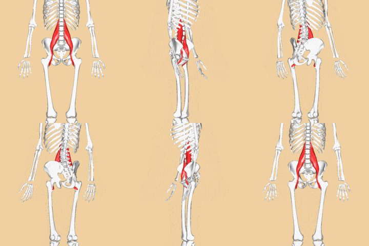
Psoas Major is a long fusiform muscle placed on the sides of the lumbar region of the vertebral column and brim of the lesser pelvis. [1] Psoas is involved in various activities of daily living like running, dancing, sitting, walking, as it is the primary connector between trunk and lower limbs.Movement at pelvis could cause changes at the lumbar spine, thereby altering the line of pull of the psoas muscle. Thus, it helps in stabilising the spine and maintaining the posture.[2]
The psoas muscles are made of both slow and fast twitch muscles fibres.The fibre type composition of the psoas major muscle indicates its dynamic and postural functions, which supports the fact that, it is the main flexor of the hip joint (dynamic function) and stabiliser of the lumbar spine, sacroiliac and hip joints (postural function). The cranial part of the psoas major muscle has a primarily postural role, whereas the caudal part of the muscle has a dynamic role.[3]
Origin[edit | edit source]
The psoas muscle is anatomically considered to have superficial and deep parts owing to the presence of branches of the lumbar plexus running through it. Superficial part - overlies the lumbar plexus and takes origin from the sides of the T12 and L1 to L4 vertebrae including the intervening intervertebral discs. The deep part which lies mainly deep to the branches of the lumbar plexus takes origin from the transverse processes of lumbar vertebrae L1 to L5.
Insertion[edit | edit source]
The fibres of the muscle converge from its wide origin as they descend on the posterior abdominal wall, They cross the pelvic inlet/brim to form a long tendon, which is joined within the pelvic region by multitudinous fibres from the iliacus muscle, finally inserting into the lesser trochanter of the femur.
Nerve Supply[edit | edit source]
Branches from the ventral rami of lumbar spinal nerves (L1, L2, and L3) before they join to form the lumbar plexus.
Blood Supply[edit | edit source]
The muscle receives blood from the four lumbar arteries from the aorta, from small branches of the renal arteries, from small muscular branches of the common iliac artery, and from the deep circumflex iliac artery.
Action
- Flexion of the thigh at the hip
- Minimal action in lateral rotation and abduction of the thigh[4]
Function[edit | edit source]
Stabilises the lumbar spine [5], maintains erect posture when working in reverse action.
Video[edit | edit source]
Clinical Importance of Psoas Muscle:[edit | edit source]
Relatively the conditions that involve psoas muscles are less common. However, a number of conditions that occurs involving iliac and psoas muscles, which includes infectious (particularly psoas abscess), haemorrhage, neoplastic lesions and injuries that take place due to trauma and during surgical procedures.[6]
Psoas Syndrome and Iliopsoas Tendinitis:[edit | edit source]
Psoas syndrome and iliopsoas tendinitis are less common musculoskeletal conditions therefore, they often go misdiagnosed. Clinically it presents symptoms like low back pain, pain in groin and pelvis, radiating pain towards knee; which halts the primary function of psoas muscle that is hip flexion (hence difficult walking) and to sustain fully erect posture. The condition is mostly found in acute/overuse injuries, more commonly sports specific like runners, athletic activities, plyometric and among ballet dancers.[7][8][9] It is known as internal coxa saltans[10].
Prolong sitting and work which requires long sitting periods, tend to put psoas in shortened and tight position leading to psoas syndrome.[11] Because they are major flexors, weak psoas muscles can cause many of the surrounding muscles to compensate and become overused. That is why a tight or overstretched psoas muscle could cause low back and pelvic pain[2]. Iliopsoas tendinitis may also cause snapping hip syndrome which is characterised by pain and snap (click) in groin.[12]
Moreover, restriction of diaphragm can also occur due to psoas dysfunction and psoas spasm can potentially cause more disability of back muscles than other pathologic condition.[13] A tight psoas muscle can create a thrusting forward of the ribcage. This causes shallow, chest breathing, which limits the amount of oxygen taken in and encourages over usage of your neck muscles[2].
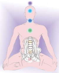
'Psoas, “the muscle of soul” [edit | edit source]
The unique location of psoas makes it the deepest core muscle of human body and the only muscle that connects trunk to lower limb. Therefore, it plays a key role in stability and balance of human body.[14] The psoas muscles support internal organs and work like hydraulic pump allowing blood and lymph to be pushed in and out of the cells.It creates a muscular shelf on which the kidneys and adrenals rest. The movement of diaphragm, during breathing gently massages the psoas muscles and neighbouring organs,thereby stimulating blood circulation. But, when the psoas muscles become imbalanced, kidneys and adrenal glands also get affected, resulting into physical and emotional exhaustion[2].
So, psoas may seem like small muscle, but its pivotal role in walking, postural alignment, balance, flexibility, joint mobility and organ functioning, makes it, clinically important, during assessment and screening.
Related links[edit | edit source]
References[edit | edit source]
- ↑ Gray, Henry. Anatomy of the Human Body. Philadelphia: Lea Febiger, 1918; Bartleby.com, 2000. www.bartleby.com/107/.
- ↑ 2.0 2.1 2.2 2.3 https://www.drnorthrup.com/psoas-muscle-vital-muscle-body/
- ↑ Arbanas J, Starcevic Klasan G, Nikolic M, Jerkovic R, Miljanovic I, Malnar D. Fibre type composition of the human psoas major muscle with regard to the level of its origin. https://www.ncbi.nlm.nih.gov/pmc/articles/PMC2796786/Journal of anatomy. 2009 Dec 1;215(6):636-41.
- ↑ http://www.wheelessonline.com/ortho/psoas
- ↑ Sandy Sajko, Kent Stuber. Psoas Major: a case report and review of its anatomy, biomechanics, and clinical implications. J Can Chiropr Assoc. 2009 Dec; 53(4): 311–318.
- ↑ Tonolini M, Campari A, Bianco R. Common and Unusual Diseases Involving the Iliopsoas Muscle Compartment: spectrum of cross-sectional imaging findings. Abdom Imaging. 2012; 37(1).
- ↑ Tufo A, Desai GJ, Cox WJ. Psoas syndrome: a frequently missed diagnosis. J Am Osteopath Assoc. 2012 Aug;112(8):522-8
- ↑ Psoas Syndrome. http://www.my.clevelandclinic.org/ (accessed 15 December 2016)
- ↑ Garry JP. Iliopsoas Tendinitis. http://www.emedicine.medscape.com/ (accessed 15 December 2016).
- ↑ Tyler TF, Fukunaga T, Gellert J. Rehabilitation of soft tissue injuries of the hip and pelvis.https://www.ncbi.nlm.nih.gov/pmc/articles/PMC4223288/ International journal of sports physical therapy. 2014 Nov;9(6):785.
- ↑ O'Dwyer S. Iliopsoas Syndrome The Hidden Root of Pain. http://www.lower-back-pain-answers.com/ (accessed 15 December 2016)
- ↑ Iliopsoas Tendonitis and Snapping Hip. https://www.health.ucsd.edu (accessed 20 December 2016)
- ↑ Tufo A, Desai GJ, Cox WJ. Psoas syndrome: a frequently missed diagnosis. J Am Osteopath Assoc. 2012 Aug;112(8):522-8
- ↑ Wilbanks B. The ‘Muscle of the Soul’ may be Triggering Your Fear and Anxiety. http://www.wakingtimes.com/ (accessed 18 December 2016


