Plantar Fasciitis
Original Editor - Brooke Kennedy
Top Contributors - Admin, Kris Porter, Rachael Lowe, Elien Lebuf, Bert Pluym, Esraa Mohamed Abdullzaher, Kim Jackson, Jonathan Wong, Chrysolite Jyothi Kommu, Brooke Kennedy, Lucinda hampton, Scott Buxton, Olajumoke Ogunleye, Jeroen Van Cutsem, Thomas Janicky, Elke Lathouwers, Vidya Acharya, Lisa Couck, Aminat Abolade, Kai A. Sigel, Tony Lowe, Khloud Shreif, Simisola Ajeyalemi, Jessica Galasso, Sehriban Ozmen, Yahya Al-Razi, Shaimaa Eldib, Saud Alghamdi, Claire Knott, Wanda van Niekerk, David Csepe, Jess Bell, Jarapla Srinivas Nayak, Keta Parikh, Stijn Van de Vondel, Habibu Salisu Badamasi, WikiSysop, Rishika Babburu and Padraig O Beaglaoich
Definition/Description[edit | edit source]
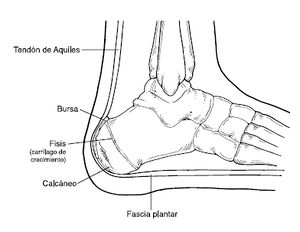
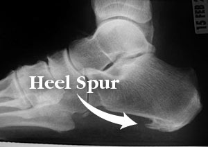
Plantar fasciitis is the result of collagen degeneration of the plantar fascia at the origin, the calcaneal tuberosity of the heel as well as the surrounding perifascial structures.[1]
- The plantar fascia plays an important role in the normal biomechanics of the foot.
- The fascia itself is important in providing support for the arch and providing shock absorption.
- Despite containing "itis," this condition is characterized by an absence of inflammatory cells, hence it is considered degenerative, and not an inflammatory pathology[2][1]. As such, “fasciosis” or “fasciopathy” are increasingly used to refer to this condition[3].
The pathology is characterized by medial heel pain that worsens with weight-bearing, as well as after rest or non-weight bearing[4]. Plantar fasciitis often presents chronically with symptoms lasting over a year in duration[5].
There are many different sources of pain in the plantar heel besides the plantar fascia and therefore the term "Plantar Heel Pain" serves best to include a broader perspective when discussing this and related pathology.
Anatomy[edit | edit source]
The plantar fascia:
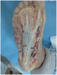
- Comprised of white longitudinally organized fibrous connective tissue which originates on the periosteum of the medial calcaneal tubercle, where it is thinner but it extends into a thicker central portion.
- The thicker central portion of the plantar fascia then extends into five bands surrounding the flexor tendons as it passes all 5 metatarsal heads.
- Pain in the plantar fascia can be insertional and/or non-insertional and may involve the larger central band, but may also include the medial and lateral band of the plantar fascia.
- Blends with the paratenon of the Achilles tendon, the intrinsic foot musculature, skin, and subcutaneous tissue.[6][7]
- This thick elastic multilobular fat pad is responsible for absorbing up to 110% of body weight during walking and 250% during running and deforms most during barefoot walking vs. shod walking.[8]
During weight-bearing:
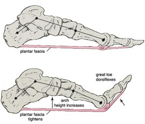
- Tibia loads the foot “truss” and creates tension through the plantar fascia (windlass mechanism see R).
- The tension created in the plantar fascia adds critical stability to a loaded foot with minimal muscle activity.[9][10][11]
Etiology[edit | edit source]
Often presents as an overuse injury, primarily due to repetitive strain causing micro-tears of the plantar fascia but can occur as a result of trauma or other multifactorial causes.
There are many risk factors for plantar heel pain including but not limited too:
- Reduced dorsiflexion and first metatarsophalangeal joint extension are weakly associated[12]
- Increased plantar flexion range[13]
- Pes cavus OR pes planus deformities
- Excessive foot pronation dynamically
- Impact/weight-bearing activities such as prolonged standing, running, etc
- Improper shoe fit
- Elevated BMI > kg/m2
- In the athletic population, BMI is not associated with increased plantar fasciitis risk, however, evidence suggests BMI is associated with increased risk in the non-athletic population[14].
- Diabetes Mellitus (and/or other metabolic condition)
- Leg length discrepancy
- Tightness and/or weakness of Gastrocnemius, Soleus, Tendoachilles tendon and intrinsic muscle.[15]
- Low quality evidence suggests an association between weight-bearing activities and plantar fasciitis[16].
Epidemiology[edit | edit source]
Plantar fasciitis is the most common cause of heel pain presenting in the outpatient setting.
- Most prevalent between 40 and 60 years of age and accounts for 15% of foot injuries in the general population[17].
- Incidence is higher among runners, accounting for about 17.4% of runner-related injuries[18]
- Thought to occur in about 10% of the general population
- 83% of these patients being active working adults between the ages of 25 and 65 years old
- 11% to 15% of all foot symptoms requiring professional medical care.
- May present bilaterally in a third of the cases[2].
- The average plantar heel pain episode lasts longer than 6 months and it affects up to 10-15% of the population.
- Approximately 90% of cases are treated successfully with conservative care.[19][20][21].
- Females present with plantar fasciitis slightly more commonly than males.[22]
- In the US alone, there are estimates that this disorder generates up to 2 million patient visits per year, and account for 1% of all visits to orthopedic clinics.
- Plantar heel pain is the most common foot condition treated in physical therapy clinics and accounts for up to 40% of all patients being seen in podiatric clinics.[23]
Diagnostic Procedures[edit | edit source]
Plantar fasciitis is a clinical diagnosis. It is based on patient history and physical exam.
- Patients can have local point tenderness along the antero-medial of the calcaneum, pain on the first steps, or after training.
- Plantar facia pain is especially evident upon the dorsiflexion of the patient's pedal phalanges, which further stretches the plantar fascia. Therefore, any activity that would increase the stretch of the plantar fascia, such as walking barefoot without any arch support, climbing stairs, or toe walking can worsen the pain.
- Clinical examination will take into consideration a patient's medical history, physical activity, foot pain symptoms, and more.
- The doctor may decide to use imaging modalities like radiographs, diagnostic ultrasound, and MRI.
Characteristics/Clinical Presentation[edit | edit source]
- Heel pain with first steps in the morning or after long periods of non-weight bearing
- Tenderness to the anterior medial heel
- Limited dorsiflexion and tight achilles tendon
- A limp may be present or may have a preference to toe walking
- Pain is usually worse when barefoot on hard surfaces and with stair climbing
- Many patients may have had a sudden increase in their activity level prior to the onset of symptoms
Examination[edit | edit source]
Take into consideration a patient's medical history, physical activity, and foot pain symptoms.
Look for the following:
- Pain reproduced by palpating the plantar medial calcaneal tubercle at the site of the plantar fascial insertion on the heel bone.
- Pain reproduced with passive dorsiflexion of the foot and toes.
- Windlass Test - Passive dorsiflexion of the first metatarsophalangeal joint (test to provoke symptoms at the plantar fascia by creating maximal stretch), positive test if pain is reproduced.[2] (shown in 40 second video below)
Secondary findings may include
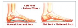
- Tight Achilles heel cord, pes planus (see R), or pes cavus.
- Altered gait (look for biomechanical factors that may predispose client plantar fascia problems) or predisposing factors mentioned previously.
- Obesity
- Work-related weight-bearing
Imaging[edit | edit source]
Ultrasonography is the most used imaging modality for this condition, and plantar fascia thickness is most often assessed - meta-analysis showed patients with plantar fasciitis have a plantar fascia 2.16 mm thicker when compared to a control group, and typically had plantar fascia thickness of 4.0 mm and above[24].
Medical Management[edit | edit source]
Conservative measures are the first choice:
- Relative rest from offending activity as guided by pain level should be prescribed.
- Ice after activity as well as oral or topical NSAIDs can be used to help alleviate pain.
- Deep friction massage of the arch and insertion.
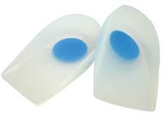
- Shoe inserts or orthotics and night splints may be prescribed in conjunction with the above.
- Educate patients on proper stretching and rehab of the: plantar fascia; achilles' tendon; gastrocnemius; and soleus.
If pain does not respond to conservative measures:
- More advanced or invasive techniques may be used eg extracorporeal shock-wave therapy [25], botulinum toxin A, autologous platelet-rich plasma, dextrose prolotherapy, or steroid injections.
- Important that advanced and invasive techniques be combined with conservative therapies.
- Surgery should be the last option if this process has become chronic and other less invasive therapies have failed[2]
Physical Therapy Management[edit | edit source]
The condition can be disabling if not appropriately managed.
An important tool is patient education:
- Patients need to be told that symptoms may take weeks or even months to improve (depending on circumstances of injury).
- To follow the advice given eg rest from aggravating activities initially, ice, stretch.
- Be aware of the importance of a home exercise plan[2]
The Clinical Practice Guidelines provide recommended physical therapy interventions based on available evidence. Interventions most recommended include: manual therapy, stretching, taping, foot orthoses, and night splints.[26]
- Manual Therapy should include soft tissue and joint mobilization.[26]
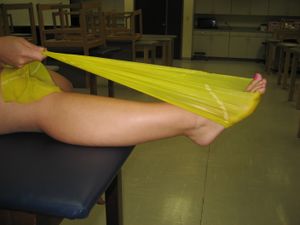
- Stretching should include the plantar fascia and gastrocnemius/Soleus complex.[26]
- Stretching the plantar fascia consists of the patient crossing the affected leg over the contralateral leg and using the fingers across the base of the toes to apply pressure into toe extension until a stretch can be felt along the plantar fascia. [27]
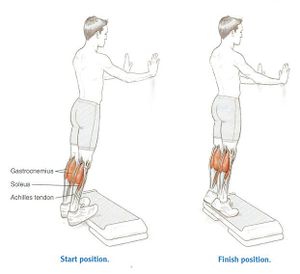
- Achilles’ tendon stretching can be performed in a standing position with the affected leg placed behind the contralateral leg with the toes pointed forward. The front knee is then bent, keeping the back knee straight and heel on the ground. The back knee could then be in a flexed position for more of a soleus stretch. According to the recent systematic review published in Life, stretching interventions seemed to improve in both pain and function over time, with a combination of calf and plantar fascia stretches was found to be most effective.[27]
- Taping should prevent pronation.[26]
- Foot orthoses can be prefabricated or custom. They must support the medial longitudinal arch and provide cushioning to the heel. [26]
- If the patient has pain with initial steps in the morning, a night splint would be beneficial. [26]
- Posterior-night splints maintain ankle dorsiflexion and toe extension, allowing for a constant stretch on the plantar fascia
According to the Clinical Practice Guidelines, ultrasound, electrotherapy, and dry needling cannot be recommended. There is some support for low-level laser, Phonophoresis with ketoprofen gel, change in footwear, weight loss, therapeutic exercise, and neuromuscular re-education.[26]
- Footwear should include a rocker-bottom shoe.[26]
- If weight is a concern, the patient should be referred to a more appropriate healthcare provider for nutritional advice.
- Therapeutic exercise and neuromuscular re-education should focus on reducing pronation and improving weight distribution in weight bearing. [26]
- Similar to tendinopathy management, high-load strength training appears to be effective in the treatment of plantar fasciitis. High-load strength training may aid in a quicker reduction in pain and improvements in function.[28] . Systematic review suggests there is minimal evidence to support the use of foot muscle training in patients with plantar fasciitis.[25]
Plantar fascia stretching video provided by Clinically Relevant
Outcome Measures[edit | edit source]
Differential Diagnosis[edit | edit source]
- Neurological - abductor digiti quinti nerve entrapment, lumbar spine disorders, problems with medial calcaneal branch of the posterior tibial nerve, tarsal tunnel syndrome
- Soft tissue - Achilles Tendinopathy, fat pad atrophy, heel contusion, plantar fascia rupture, posterior tibial tendonitis, retrocalcaneal bursitis
- Skeletal - Severs' disease, calcaneal stress fracture, infections, inflammatory arthropathies, subtalar arthritis
- Miscellaneous - metabolic disorders, osteomalacia, Paget's disease, sickle cell disease, tumors (rare), vascular insufficiency, Rheumatoid arthritis
Concluding Comments[edit | edit source]
- Thorough patient education needed.
- Usually a self-limiting condition, and with conservative therapy, symptoms are usually resolved within 12 months of initial presentation and often sooner.
- Sometimes more chronic cases of this condition will need additional follow-up to consider more advanced therapies and evaluation of gait and biomechanical factors that can potentially be corrected through gait retraining.
- Corticosteroid injections have been shown to be beneficial in the short-term (less than four weeks) but ineffective in the long term.
- Evidence of the efficacy of platelet rich plasma, dex prolotherapy, and extra-corporeal shockwave therapy is conflicting[2].
References[edit | edit source]
- ↑ 1.0 1.1 Lemont H, Ammirati KM, Usen N. Plantar fasciitis: a degenerative process (fasciosis) without inflammation. J Am Podiatr Med Assoc. 2003;93(3):234–7.
- ↑ 2.0 2.1 2.2 2.3 2.4 2.5 Buchanan BK, Kushner D. Plantar fasciitis.Available from:https://www.ncbi.nlm.nih.gov/books/NBK431073/ (last accessed 22.6.2020)
- ↑ Rhim HC, Kwon J, Park J, Borg-Stein J, Tenforde AS. A systematic review of systematic reviews on the epidemiology, evaluation, and treatment of plantar fasciitis. Life (Basel, Switzerland). 2021;11(12):1287.
- ↑ Schepsis AA, Leach RE, Gorzyca J. Plantar fasciitis : etiology, treatment, surgical results, and review of the literature. Clinical orthopaedics and related research. 1991;266(266):185–96.
- ↑ Klein SE, Dale AM, Hayes MH, Johnson JE, McCormick JJ, Racette BA. Clinical Presentation and Self-Reported Patterns of Pain and Function in Patients with Plantar Heel Pain. Foot & Ankle International. 2012;33(9):693-698.
- ↑ Carlson RE, Fleming LL, Hutton WC. The biomechanical relationship between the tendoachilles, plantar fascia and metatarsophalangeal joint dorsiflexion angle. Foot ankle Int / Am Orthop Foot Ankle Soc [and] Swiss Foot Ankle Soc. 2000;21(1):18–25.
- ↑ Stecco C, Corradin M, Macchi V, et al. Plantar fascia anatomy and its relationship with Achilles tendon and paratenon. J Anat. 2013;223(August):1–12. doi:10.1111/joa.12111.
- ↑ Gefen A, Megido-Ravid M, Itzchak Y. In vivo biomechanical behavior of the human heel pad during the stance phase of gait. J Biomech. 2001;34:1661–1665. doi:10.1016/S0021-9290(01)00143-9
- ↑ Tweed JL, Barnes MR, Allen MJ, Campbell J a. Biomechanical consequences of total plantar fasciotomy: a review of the literature. J Am Podiatr Med Assoc. 2009;99(5):422–30.
- ↑ Cheung JT-M, An K-N, Zhang M. Consequences of partial and total plantar fascia release: a finite element study. Foot ankle Int / Am Orthop Foot Ankle Soc [and] Swiss Foot Ankle Soc. 2006;27(2):125–32. Available at: http://www.ncbi.nlm.nih.gov/pubmed/16487466.
- ↑ Crary JL, Hollis JM, Manoli A. The effect of plantar fascia release on strain in the spring and long plantar ligaments. Foot ankle Int / Am Orthop Foot Ankle Soc [and] Swiss Foot Ankle Soc. 2003;24(3):245–50
- ↑ Irving DB, Cook JL, Menz HB. Factors associated with chronic plantar heel pain: a systematic review. Journal of science and medicine in sport. 2006;9(1):11–22.
- ↑ Hamstra-Wright KL, Huxel Bliven KC, Bay RC, Aydemir B. Risk Factors for Plantar Fasciitis in Physically Active Individuals: A Systematic Review and Meta-analysis. Sports Health. 2021 May-Jun;13(3):296-303. doi: 10.1177/1941738120970976. Epub 2021 Feb 3. PMID: 33530860; PMCID: PMC8083151.
- ↑ Butterworth, P.A., Landorf, K.B., Smith, S.E. and Menz, H.B. (2012), The association between body mass index and musculoskeletal foot disorders: a systematic review. Obesity Reviews, 13: 630-642. https://doi.org/10.1111/j.1467-789X.2012.00996.x
- ↑ Lemont H, Ammirati KM, Usen N. Plantar fasciitis: a degenerative process (fasciosis) without inflammation. Journal of the American Podiatric Medical Association. 2003 May;93(3):234-7.
- ↑ Waclawski ER, Beach J, Milne A, Yacyshyn E, Dryden DM. Systematic review: plantar fasciitis and prolonged weight bearing. Occup Med (Lond). 2015 Mar;65(2):97-106. doi: 10.1093/occmed/kqu177. Epub 2015 Feb 17. PMID: 25694489.
- ↑ Agyekum EK, Ma K. Heel pain: A systematic review. Chinese journal of traumatology. 2015;18(3):164–9
- ↑ Lopes AD, Hespanhol LC, Yeung SS, Costa LOP. What are the Main Running-Related Musculoskeletal Injuries? Sports medicine (Auckland). 2012;42(10):891–905.
- ↑ McPoil TG, Martin RL, Cornwall MW, Wukich DK, Irrgang JJ, Godges JJ. Heel pain--plantar fasciitis: clinical practice guildelines linked to the international classification of function, disability, and health from the orthopaedic section of the American Physical Therapy Association. J Orthop Sports Phys Ther. 2008;38(4):A1–A18. doi:10.2519/jospt.2008.0302.
- ↑ Riddle DL, Pulisic M, Pidcoe P, Johnson RE. Risk factors for Plantar fasciitis: a matched case-control study. J Bone Joint Surg Am. 2003;85-A(5):872–7
- ↑ Thomas JL, Christensen JC, Kravitz SR, et al. The diagnosis and treatment of heel pain: a clinical practice guideline-revision 2010. J Foot Ankle Surg. 2010;49(3 Suppl):S1–19. doi:10.1053/j.jfas.2010.01.001
- ↑ Lopes AD, Hespanhol Júnior LC, Yeung SS, Costa LOP. What are the main running-related musculoskeletal injuries? A Systematic Review. Sports Med. 2012;42(10):891–905. doi:10.2165/11631170-000000000-00000.
- ↑ 2002 Podiatric Practice Survey. Statistical results. J Am Podiatr Med Assoc. 2003;93(1):67–86. Available at: http://www.ncbi.nlm.nih.gov/pubmed/12533562.
- ↑ Rhim HC, Kwon J, Park J, Borg-Stein J, Tenforde AS. A Systematic Review of Systematic Reviews on the Epidemiology, Evaluation, and Treatment of Plantar Fasciitis. Life [Internet]. 2021 Nov 24;11(12):1287. Available from: http://dx.doi.org/10.3390/life11121287
- ↑ 25.0 25.1 Rhim HC, Kwon J, Park J, Borg-Stein J, Tenforde AS. A Systematic Review of Systematic Reviews on the Epidemiology, Evaluation, and Treatment of Plantar Fasciitis. Life. 2021 Dec;11(12):1287.
- ↑ 26.0 26.1 26.2 26.3 26.4 26.5 26.6 26.7 26.8 J Orthop Sports Phys Ther 2014;44(11):A1–A23. doi:10.2519/jospt.2014.0303
- ↑ 27.0 27.1 DioGiovanni BF, Nawoczenski DA, Lintal ME et al. Tissue-specific plantar fascia-stretching exercise enhance outcomes in patients with chronic heel pain. Journal of Bone and Joint Surgery. 2003;85-A:1270-1277. (level of evidence: 1b)
- ↑ Rathleff, M.S., Mølgaard, C.M., Fredberg, U., Kaalund, S., Andersen, K.B., Jensen, T.T., Aaskov, S. and Olesen, J.L., 2015. High‐load strength training improves outcome in patients with plantar fasciitis: A randomized controlled trial with 12‐month follow‐up. Scandinavian journal of medicine & science in sports, 25(3). (level of evidence: 1b)
