Pectineus Muscle
Description[edit | edit source]
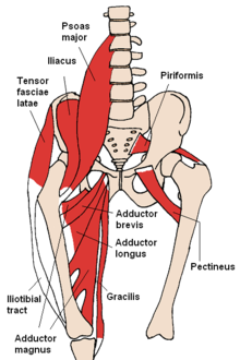
is a flat, quadrangular muscle, situated at the anterior part of the upper and medial aspect of the thigh. The pectineus muscle is the most anterior dductor of the hip [1].
It can be classified in the medial compartment of thigh(when the function is emphasized) or the anterior compartment of thigh (when the nerve is emphasized)[1].
Origin[edit | edit source]
It has the most superior attachment of all the thigh adductors, originating from the pectineal line of pubis on the superior pubic ramus. The muscle then slides over the superior margin of superior pubic ramus and courses posterolaterally down the thigh, sometimes being partially divided into a larger anterior (superficial) layer and smaller posterior (deep) layer. The layers are innervated by different nerves[2].
Insetion[edit | edit source]
Pectineus muscle inserts into the posterior surface of femur, along the pectineal line and proximal part of linea aspera[2].
Nerve supply[edit | edit source]
Pectineus is predominately innervated by the femoral nerve (L2, L3)]. However, in some people pectineus may receive innervation from two separate nerves of the lumber plexus[3].
This composite innervation reflects the dual compartmentalisation of pectineus into both the anterior and medial compartments of the thigh. In these cases the anterior part of the muscle sits is innervated by the femoral nerve (L2, L3), a feature of muscles of the anterior thigh. While the posterior, smaller part of the muscle is supplied by a branch of obturator nerve (L2, L3), the accessory obturator nerve[3].
Blood supply[edit | edit source]
The superficial part of the muscle is supplied by the medial circumflex femoral artery, a branch of the femoral artery. Deep portion of the muscle is vascularised by the anterior branch of obturator artery, itself a branch of the internal iliac artery.
Relation[edit | edit source]
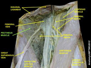
The muscle lies in the frontal plane and medially to,adductor longus. While laterally, it is related to the psoas major muscle and the medial circumflex femoral artery and vein.
The anterior surface of pectineus forms the medial part of the floor of femoral traingle together with adductor longus.
This surface of pectineus is covered with the deep layer of fascia lata that separates it from the femoral artery, femoral vein and great saphenous vein that course through the femoral triangle.
Posterior to pectineus are the adductor magnus, adductor brevis and obturator externus muscles, and the anterior branch of obturator nerve.[2][3]
Action[edit | edit source]
Due to the course of its fibers, pectineus both flexes and adducts the thigh at the hip joint when it contracts. When the lower limb is in the anatomical position, contraction of the muscle first causes flexion to occur at the hip joint. This flexion can go as far as the thigh being at a 45 degree angle to the hip joint[4].
At that point, the angulation of the fibers is such that the contracted muscle fibers now pull the thigh towards the midline, producing thigh adduction[4].
Injury[edit | edit source]
The pectineus muscle can become injured by overstretching; specifically, by stretching a leg or legs too far out to the side or front of the body. Pectineus injuries can also be caused by rapid movements like kicking or sprinting, changing directions too quickly while running, or even by sitting with a leg crossed for too long.[3]
Symptoms of the injury[edit | edit source]
The most common symptom of an injured pectineus muscle is pain. Other may include bruising,swelling,tenderness and stiffness[2].
Treatment[edit | edit source]
Treatment of pectinus muscle injury is ask patient to protect the injured from movements and objects that might cause further injury; minimize activities that use the pectinus muscle, like walking and running, in order to allow the muscle time to heal; and ice the injury to decrease and prevent swelling, as well as help decrease any pain.icing shouhd be performed 15:20 minutes every few hours[2].
References[edit | edit source]
- ↑ 1.0 1.1 Mosby's Medical, Nursing & Allied Health Dictionary, Fourth Edition, ., 1994
- ↑ 2.0 2.1 2.2 2.3 2.4 Moore,. .Clinically Oriented Anatomy (7th ed). (2014)
- ↑ 3.0 3.1 3.2 3.3 Standring,. Gray's Antomy (41tst ed.). (2016)
- ↑ 4.0 4.1 Palastanga,. . Anatomy and human movement: structure and function (6th ed.).(2012)


