Orthotics in Cerebral Palsy
Introduction[edit | edit source]
Why are the interdisciplinary team members convinced to use the orthoses as part of the treatment plan? Because of the comprehensive understanding of the CP patients, concentrating on the function limitations has a great effect on the new range of improved designs of orthoses to improve the outcome for the benefit of the patient.
In 1994 during the consensus conference held in Duke University, ISPO (International Society for Prosthetics and Orthotics) identifies the goals of the lower limb orthotic management of CP. The identified goals can also be applied in postural impairments of the trunk and upper limbs.
- To correct and/or prevent deformity
- To provide a base of support
- To facilitate training in skills
- To improve the efficiency of gait
It is important that the interdisciplinary team check the patient’s functional limitations according to the GMFCS in order to plan the treatment. The type and design of the orthosis is decided accordingly and can be changed periodically depending on the improvement of the patient condition.
Types of Orthotics[edit | edit source]
Under the International Standard terminology, orthoses are classified by an acronym describing the anatomical joints which they contain. For example, an ankle foot orthosis ('AFO') is applied to the foot and ankle, a thoracolumbosacral orthosis ('TLSO') affects the thoracic, lumbar and sacral regions of the spine. It is also useful to describe the function of the orthosis.
Types of orthoses which can be used for individuals with Cerebral Palsy are shown in the short video below then described in greater detail in the text that follows.
Lower Limb Orthotic Designs[edit | edit source]
Foot Orthoses (FO)[edit | edit source]
Foot orthotics do not prevent deformity. They provide a better contact of the sole of the foot with the ground.
Supramalleoler Orthosis (SMO)[edit | edit source]
This orthosis extends to just above the malleoli and to the toes. Consider in mild dynamic equinus, varus and valgus instability.
University of California Biomechanics Laboratory Orthosis (UCBL)
The medial side is higher than the lateral, holds the calcaneus more firmly, supports the longitudinal arch. Prescribed for hind and midfoot instability.
Ankle Foot Orthoses (AFO)[edit | edit source]
The AFO is the basic orthosis in CP and is a crucial piece of equipment for many children with spastic diplegia. The main function of the AFO is to maintain the foot in a plantigrade position. This provides a stable base of support that facilitates the function and also reduces tone in the stance phase of the gait. The AFO supports the foot and prevents drop foot during swing phase. If worn at night, a rigid AFO may prevent contracture. AFOs provide a more energy efficient gait. The brace should be simple, light but strong. It should be easy to use. Most importantly it should provide and increase functional independence.
There are various types of the AFO.
Dynamic AFO (DAFO)[edit | edit source]
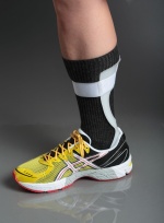
The Dynamic Ankle Foot Orthosis generally refers to a custom made Supra Malleolar Orthosis fabricated from thin thermoplastic material (FIGURE 1). It fits the foot intimately and the use of the flexible and thin thermoplastic means that the DAFO can provide circumferential control of the rear and fore foot to maintain a neutral alignment. In the original designs of DAFOs, a ‘neurological’ foot plate was often incorporated that consisted of a pad at the peronal notch, a metatarsal dome and dorsiflexing the lateral four toes. It was theorised that applying these pressures to the foot would decrease the level of spasticity in the gastrocnemius and therefore reduce the level of equinus often observed in spastic CP gait. However current literature shows there is no reduction in EMG activity of gastrocnemius with these modifications and therefore no reduction in muscle tone and the associated equinus. To effectively control sagittal plane deformities such as a plantar flexed ankle, a long lever arm is required that involves extending the trim lines up to the proximal calf. Therefore, DAFOs should only be used where there is coronal or transverse plane deformities of the foot and ankle that can be passively corrected with minimal force.
Solid AFO [edit | edit source]
The solid or rigid AFO allows no ankle motion, it covers the back of the leg completely and extends from just below the fibular head to metatarsal heads. The solid AFO enables heel strike in the stance phase and toe clearance in the swing phase. It can improve knee stability in ambulatory children. It also provides control of varus/ valgus deformity. Solid AFOs provides ankle stability in the standing frame in non-ambulatory children.
A solid ankle foot orthosis aims to prevent all movement of the foot and ankle at the talo-crural, subtalar and midfoot joints. It is prescribed to children with CP when there is:
- Moderate to high tone in the gastrocnemius muscle;
- Less than 10 degrees of ankle dorsiflexion with the knee in maximum extension,
- Moderate to severe medio lateral instabilities at the ankle
- A requirement to provide proximal control at the knee and hip joints.
It is crucial that a solid AFO is sufficiently stiff at the ankle and does not flex or ‘buckle’ during mid to late stance as the dorsiflexion moment is applied. Flexing at the ankle compromises the midfoot control a Solid AFO can provide and reduces the influence it can have at the proximal joints of the hip and knee.
A Solid AFO may be prescribed to help reduce the effects of ‘crouch’ gait, where the hips and knees are in a flexed position during mid stance. If the solid AFO does not resist the dorsiflexion moment during mid to late stance, the tibial Shank to Vertical Angle (SVA) is inclined and the Ground Reaction Force is shifted posterior at the knee and anterior to the hip, thereby permitting crouch gait to occur.
The ankle section of solid AFO may be stiffened by:
- Selecting a material that is sufficiently stiff with an appropriate thickness to manufacture the AFO;
- Ensuring the trimlines/edges at the ankle section are anterior to the malleoli;
- Adding reinforcement material by ‘double moulding’ thermoplastic, including ribbing at the ankle or using carbon fibre reinforcements.
Posterior Leaf Spring AFO (PLSO)[edit | edit source]
A Posterior Leaf Spring AFO is a rigid AFO trimmed behind the malleoli’s to provide flexibility at the ankle and allows passive ankle dorsiflexion during the stance phase. A PLSO provides smoother knee-ankle motion during walking while preventing excessive ankle dorsiflexion Varus-valgus control is also poor because it is repeatedly deformed during weight bearing. A PLSO is an ideal choice in mild spastic equinus. Do not use it with patients who have crouch gait and pes valgus.
The Posterior Leaf Spring (PLS) AFO is deemed a swing phase orthosis in that it is effective during swing phase only. It is only suitable for children who present with isolated dorsiflexor weakness or paralysis. It may be manufactured from many different types of materials including Ortholen, Co-polymer polypropylene and carbon composites. The flexible nature of the PLS AFO and the elastic properties of the materials used to fabricate it produces a dynamic orthosis. It permits controlled plantarflexion in early stance phase during loading of the limb and then maintains the foot at plantargrade during swing phase to ensure the foot clears the ground. It is not able to provide adequate control of the foot and ankle in the presence of moderate to high spasticity, mediolateral instabilities at the foot or ankle or where stance phase control of the knee or hip is required. The orthotic treatment goal of the PLS AFO is to maintain the foot and ankle in a plantargrade position during swing to permit foot clearance, but permit ankle plantarflexion and dorsiflexion during stance phase.
Ground Reaction AFO (GRAFO)[edit | edit source]
This AFO is made with a solid ankle, the upper portion wraps around the anterior part of the tibia proximally with a solid front over the tibia. The rigid front provide strong ground reaction support for patients with weak triceps surae. The foot plate extends to the toes. The ankle may be set in slight plantar flexion of (2-3 degrees) if more corrective force at the knee is necessary. Use the GRAFO in patients with quadriceps weakness or crouch gait. It is an excellent brace for patients with weak triceps surae following hamstring lengthening. Children with static or dynamic knee flexion contractures (more than 15 degrees) do not get benefit out of it and do not tolerate the GRAFO.
The Ground Reaction Ankle Foot Orthosis (GRAFO is a type of solid AFO with the primary aim of increasing knee control during stance phase. The GRAFO generally has an anterior pre-tibial shell to increase the proximal lever arm and help control tibial progression through stance phase. As with the solid AFO it is imperative the GRAFO is sufficiently stiff to resist the dorsiflexion moment during mid-late stance phase to ensure it can help influence the position of the Ground Reaction Force in relation to the knee and hip joints. A full length footplate should be used in the GRAFO design to provide the maximum foot lever length thus shifting the GRF as far anterior to the knee as possible during stance phase. It is also important to ensure this footplate is sufficiently stiff to resist dorsiflexion during late stance. This can be achieved by ensuring the footplate material is sufficiently stiff and thick, but also by extending the medial and lateral trim lines distally to cover the metatarsal phalangeal joints. Either dynamic (tone) or fixed (contracture) hip or knee flexion contractures of >10 degrees or transverse plane deformities such as excessive femoral and tibial torsion will reduce the effectiveness of the GRAFO at the knee and hip joints due to reduced foot lever length. To ensure optimal function, it is imperative that the GRAFO is aligned or ‘tuned’ to ensure the GRF is anterior to the knee at mid-late stance to help generate a knee extension moment.
Anti-Recurvatum AFO
This special AFO is molded in slight dorsiflexion or has the heel built up slightly to push the tibia forward to prevent hyperextension during stance phase. Consider prescribing this AFO for the treatment of genu recurvatum in hemiplegic or diplegic children. Anti-recurvatum AFOs may be solid or hinged depending on the child’s tolerance.
Hinged AFO[edit | edit source]
Hinged AFOs have a mechanical ankle joint usually preventing plantar flexion, but allowing relatively full dorsiflexion during the stance phase of gait. They provide a more normal gait because they permit dorsiflexion in stance phase of the gait, thus making it easier to walk on uneven surfaces and stairs. This is the best AFO for most ambulatory patients. Adjust the plantar flexion stop in (3- 7 degrees) dorsiflexion to control knee hyperextension in stance in children with genu recurvatum. The hinged AFO is contraindicated in children who do not have passive dorsiflexion of the ankle because it may force the midfoot joints into dorsiflexion and cause midfoot break deformity. Knee flexion contractures and triceps weakness are other contraindications where a hinged AFO may increase crouch gait. The AFO may be fitted with a hinge that allows 10 degrees passive dorsiflexion while preventing plantar flexion. This creates a more natural gait.
Hinged AFOs incorporate a mechanical joint that either allows or assists motion in one or more directions. Typically, in children with CP, hinged AFOs prevent plantarflexion at plantargrade (90degrees) and then permit free dorsiflexion. This design of AFO should only be considered if there is sufficient gastrocnemius length that permits 10degrees of dorsiflexion with the knee in full extension and where there is no spastic catch or resistance in range of the gastrocnemius due to increased muscle tone. Any AFO that permits the ankle to be in more dorsiflexion than can be achieved with the knee in maximum extension, will actually limit knee extension in stance and adversely affect knee and hip kinetics .The hinged AFO should also be only used where there is sufficient control of knee joint flexion and no requirement to prevent knee flexion in stance phase. Permitting ankle dorsiflexion in this case, shifts the GRF posterior to the knee and causes a knee flexion moment. Even if there is sufficient gastrocnemius ROM and knee control, hinged AFOs may be unsuitable in the presence of moderate to severe medio-lateral instabilities of the foot and ankle.
Knee Orthoses
Knee orthoses are used as resting splints in the early postoperative period and during therapeutic ambulation. There are two types of knee orthoses, the knee immobilizer and the plastic knee-ankle foot orthosis (KAFO). The use of such splints protects the knee joint, prevents deformity recurrence after multilevel lengthening and enables a safer start to weight bearing and ambulation after surgery.
Knee Immobilizers[edit | edit source]
Knee immobilizers are made of soft elastic material and hold only the knee joint in extension, leaving the ankle joint free. Consider using them in the early postoperative period after hamstring surgery and rectus tendon transfers.
Consider the knee immobilizer after hamstring surgery.
Plastic KAFOs[edit | edit source]
Plastic resting KAFOs extend from below the hips to the toes and stabilize the ankle joint as well as the knee. They are more rigid and provide better support to the ankle and the knee in the early postoperative phase. Knee-ankle-foot orthoses with metal uprights and hinged joints (KAFOs) were developed and used extensively in the 1950s and 60s for children with poliomyelitis. Though KAFOs are still used for ambulation in poliomyelitis and myelomeningocele where there is a need to lock the knee joint, they are not useful for the child with CP because they disturb the gait pattern by locking the knee in extension in the swing phase. Donning the KAFO on and off takes a lot of time and they are difficult to wear. For these reasons, KAFOs for functional ambulation have disappeared from use in children with CP. Use anti recurvatum AFOs or GRAFOs for knee problems in ambulatory children.
Use the plastic KAFO at night and in the early postoperative period after
Multi-level surgery to protect the extremity while allowing early mobilization.
Care of Orthosis[edit | edit source]
It is important that the orthosis is in good working order and shows minimal signs of wear and tear. Check that components are in good condition and if hinged check they are functioning and locking if needed. Check the fit also and teach client in hygiene and care aspects of orthosis. See video below.
Hip Abduction Orthoses[edit | edit source]
Consider using hip abduction orthoses in children with hip adductor tightness to protect hip range of motion and prevent the development of subluxation. One clear indication for hip abduction orthoses is the early period after adductor lengthening.
Spinal Orthotics[edit | edit source]
There are various types of braces used for spinal deformity. This braces are not prescribed in order to stop the progression of scoliosis but to provide better sitting balance. As most children with scoliosis need spinal surgery to establish and maintain sitting balance in the long run. A thoraco-lumbo-sacral brace helps the child to sit better during the growth spurt period when spinal deformity becomes apparent, progresses fast and the child out grows custom molded seating devices quickly. Children who are not candidates for surgery for different reasons may use spinal braces instead of seating devices for better sitting.
Upper Limb Orthotics[edit | edit source]
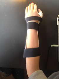
The indications of bracing in the shoulder and elbow are very limited. An example of a resting splint is a thermoplastic resting wrist and hand splint which keeps the wrist in 10-20 degrees extension, the metacarpal phalangeal joint(MPJ) in 60 degrees flexion and the interphalangeal joint (IPJ) in extension ( see figure on R). This type of splint is used at night and during periods of inactivity with the hope of preventing deformity. An example of a functional splint is an opponents splint, which can be used in everyday activities. Hand orthoses may inhibit the active use of the extremity and effect sensation of the hand in a negative way. Use them only in the therapy setting or at school and take them off during other times in the day.
These are the most known type of orthoses used in one stage of the Cerebral Palsy Treatment Plan, bearing in mind with Cerebral Palsy a periodical orthosis assessment has to be done in order to decide if there is a need for changing the design or type.
Orthotic Prescription[edit | edit source]
Lower Limb Orthoses for Ambulatory Children (GMFCS I, II and III)[edit | edit source]
Ambulatory children with Cerebral Palsy often present with numerous gait deviations that primarily result from the loss of selective motor control, decreased muscle strength and abnormal muscle tone [3]. These motor disorders of Cerebral Palsy are frequently accompanied by disturbances of sensation, perception, cognition, communication, behaviour and epilepsy [4]. Hence, the orthotic management of ambulatory children with Cerebral Palsy requires comprehensive rehabilitation using age-appropriate interventions that encompass the ICF domains of body function and structure, activity and participation, personal and environmental factors [5]. Orthoses are used to manage the secondary musculoskeletal problems of muscle contracture and bony deformity. Without appropriate orthotic intervention, detrimental changes to the gait and function of the child with Cerebral Palsy will occur over less than two years [6].
A diagnosis of CP does not correlate with any clearly defined rehabilitative intervention strategies, nor does it correlate with a defined set of expected outcomes for the child and family [7]. To provide effective orthotic intervention for children with Cerebral Palsy it is important to clearly identify the functional abilities of each child in order to establish the aims of any orthotic intervention. Through a consensus conference in 1994, the International Society of Prosthetics and Orthotics (ISPO) identified the aims of lower limb orthotic management of cerebral palsy as:
- To correct and/or prevent deformity.
- To provide a base of support.
- To facilitate training in skills.
- To improve the efficiency of gait [8]
A literature review conducted by Figuerdo et. al [9] and a report from the ISPO Cerebral Palsy Consensus Conference of 2008 [10]criticised the evidence base relating to the orthotic management of children with Cerebral Palsy. Both documents identified a relatively low amount of research that dealt specifically with the orthotic management of children with Cerebral Palsy.
They also found many of these studies employed poor methodologies causing the evidence to be of a lower scientific quality. Hence, it was recommended in both the literature review and the consensus conference that future studies have more robust methodologies and provide more in-depth descriptions of the participant presentations, the methods used and the orthotic interventions provided [11]. This will allow the results from future studies to be transferrable to clinical practice.
The incomplete reporting of orthoses in the scientific literature was highlighted as a major area of concern in the literature review and the consensus report. Many studies evaluating the efficacy of orthotic intervention in children with Cerebral Palsy simply described the orthosis being tested as an ‘AFO’. Without sufficient details on the construction material, trim lines used and alignment of the AFOs, it is impossible to replicate the orthosis and allow an orthotist to transfer the orthotic design to their own clinical practice. Ridgewell et produced a systematic literature review to evaluate the level and quality of detail reported about participants, devices and testing protocols to generate best practice guidelines for reporting of orthoses in future studies examining children with Cerebral Palsy. They reiterated that many of the papers failed to provide sufficient information that could allow the synthesis of the information to contribute to the orthotic evidence base.
The body of knowledge on the efficacy of AFOs will gradually grow using well designed studies and provided homogenous patient groups are measured, relevant outcome measures are used and the AFOs evaluated in the study are unambiguously mechanically characterised [12].
Despite some shortcomings in the current literature, there is sufficient evidence available to establish 4 key points on the efficacy of the orthotic management of children with Cerebral Palsy:
- AFOs provide positive influences on the temporal spatial characteristics, kinematics and kinetics of gait in children with Cerebral Palsy.
- AFOs can reduce metabolic cost and the energy expenditure of walking.
- AFOs provide positive effects on ability and function.
- ‘Tuning’ the AFO and footwear combination (AFO-FC) is critical to optimise the biomechanical benefits of the orthosis and enable positive influences on the knee and hip joints.
Lower Limb Orthoses for Non-Ambulatory Children (Pre-standing and GMFCS IV and V)[edit | edit source]
Hip Instability (GMFCS IV and V)[edit | edit source]
Hip subluxation and dislocation due to spasticity is the second most common musculoskeletal deformity seen in children with CP. The GMFCS level of the child is strongly associated with hip displacement, as the lower levels of motor function have increased predictive rates of hip displacement. The overall incidence has been described in the literature at around 35% with variances of around 1% of children with spastic hemiplegia affected up to 75% of children with spastic quadriplegia [13]. Hip displacement that leads to subluxation is associated with greater functional activity limitations, increased pain, development of pelvic obliquity and in turn progressive scoliosis.
There is no evidence that hip abduction orthoses prevent progressive hip displacement over time. A randomized control trial monitored children over one year and compared the use of botulinum toxin type A (BoNT-A) to the adductors and hamstrings and a variable hip abduction orthosis (SWASH) with a control group that received physiotherapy but no orthoses on gross motor function, hip displacement and surgery rates. There were no significant differences between the groups at one year follow up in either the control group or those that received BoNT-A and a hip abduction orthosis. A long term follow-up of three years to this original study found that BoNT-A and hip abduction bracing does not reduce the need for surgery or improve hip development at skeletal maturity [14].
While hip abduction orthoses cannot prevent the development and progression of hip displacement and subluxation, they may improve sitting posture, symmetry and comfort in non-ambulant children. Ambulant children may also gain some benefit from hip orthoses that control adduction by decreasing the effects of a scissoring gait, leading to increased standing stability and gait efficiency. There is little evidence to support the widespread provision of a hip abduction orthoses for children with CP. The prescription of hip orthoses for both ambulant and non-ambulant children must be on a case by case basis. The prescription must be intrinsically linked to pre-determined rehabilitation goals and objectively assessed with appropriate outcome measures. If it is found that the hip abduction orthosis is not achieving the rehabilitation goals wear should be stopped.
Ankle Foot Orthoses (GMFCS IV and V)[edit | edit source]
Children in GMFCS levels IV and V will spend a large amount of their time in seated positions, meaning they are more likely to develop flexion contractures. It has been found that maintaining a spastic muscle in maximum extension for 6-8 hours can help to reduce the development of flexion contractures [15]. AFOs for this group of children are often prescribed to manage the resultant equinus deformity at the foot and ankle with the functional goals of ensuring the child is able to use a standing frame and perform assisted standing transfers if appropriate. However, the gastrocnemius muscle is a bi-articulate muscle, meaning that it crosses both the knee and ankle joints. To provide an appropriate stretch of the gastrocnemius muscle, it is crucial the knee is held in maximum extension with the foot and ankle in maximum dorsiflexion. Therefore, AFOs must be combined with another orthosis such as a stiffened fabric gaiter or a 3 point knee brace to ensure the gastrocnemius is stretched. AFOs may also be used to manage coronal and transverse plane deformities of the foot in children with GMFCS levels IV and V. Mobile deformities including rear foot varus/valgus and forefoot abduction/adduction and supination/pronation, may be corrected in the casting process and controlled using solid AFOs. Any fixed deformity must be accommodated and maintained in their ‘best’ corrected or most neutral position.
Spinal Orthoses (GMFCS IV and V)[edit | edit source]
Children with CP who are more limited in their functional ability are at a greater risk of developing combinations of scoliosis, lordosis and kyphosis. The more severe the deficit, the more likely is spinal deformity to occur, the earlier the age of onset, and curves are likely to be more severe. The progression of the curve becomes more apparent during spinal growth and will continue into adult life. In general, if a child with CP is able to walk, then the chances of developing a severe scoliosis is much less likely compared with wheelchair dependent children. Scoliosis in children with CP has been linked to the effects of gravity when they are placed in a seated position for long periods of time [16].
The use of spinal orthoses is first line treatment for children with CP who have a related spinal deformity. There is a little evidence to support the use of spinal bracing and provision is on an individual case basis. Any prescription of a spinal orthosis must be combined with the use of seating and sleep systems and also include the use of a standing frame and/or orthoses to help reduce the effects of gravity on the spine and digestive system in the seated position.
Rigid thermoplastic spinal braces (Thoraco Lumbar Spinal Orthoses similar to those used to manage idiopathic scoliosis, are often not well tolerated by children with Cerebral Palsy as there has been reports of reduced tolerance due to pressure sores and skin irritation [17]. TLSOs that are made of more flexible material such as polyethelyne, have been found to be better tolerated and provide improved head and trunk control, improved postural position and increased sitting stability [18] [19]. Although spinal curves appear to progress in most non-ambulant children with Cerebral Palsy, there is a small cohort fitted with a semi-rigid TLSO that experience either slowing of the rate of progression or halting of curve progression. It appears that the more flexible curves respond better to orthotic intervention and this is a good predictor for prescription of a spinal orthosis.
Orthotic Management[edit | edit source]
Lower Limb Transverse Plane and Coronal Plane Deformities[edit | edit source]
The identification of the primary cause of lower limb transverse and coronal plane deformities is imperative to selecting appropriate orthotic interventions. Leg Length Discrepancies (LLDs) are common in children with hemiplegia due to Developmental Dysplasia of the Hip (DDH) and/or uneven loading of the limbs leading to decreased bone growth. The resultant pelvic obliquity and functional deficits of contralateral hip adduction and ipsilateral hip abduction can be addressed by using a shoe raise.
A ‘scissor’ gait or ‘in-toeing’ gait are commonly observed gait deviations in the presence of torsional deformities of the lower limb. In this instance it is crucial that a rotational profile of the lower limbs is performed. This will permit the orthotist to determine if the torsional deformity is of bony or muscular origin. Femoral and tibial rotational deformities and any fixed muscle contracture require surgical correction and cannot be managed with orthoses. Orthoses that have been used to manage torsional deformities largely rely upon derotating the affected limb through the use of ‘twister cables’ within a Hip Knee Ankle Foot Orthosis (HKAFO) or through the use of fabric garments. It is not recommended to provide a derotational orthosis that crosses the knee joint, as the applied torque leads to excessive strain on the soft tissues of the knee joint.
Tuning the Ankle Foot Orthosis and Footwear Combination[edit | edit source]
For an energy efficient gait, good muscular control is required to ensure appropriate alignment of the GRF relative to the ankle, knee and hip joints throughout stance phase.In the pathological gait of a child with CP, adequate control of the
GRF is not always possible due to the compromised neuromuscular system. AFOs are often prescribed to assist with lower limb control in children with CP as they have been shown to positively influence the kinetics and kinematics of gait.
To achieve optimal knee extension during the stance phase of gait, there is a common misunderstanding that the talo-cural joint must be positioned at 90° and the tibial and thigh shanks vertically aligned.This has lead to the misconception that the AAAFO should be set at 90° and that dorsi flexed or plantar grade AFOs are acceptable but that plantar flexed AFOs are not.[3] [5]
The Shank to Vertical Angle (SVA) is defined as the angle of the tibial shank relative to the vertical in the sagittal plane and may be described in degrees of incline or recline from the vertical.[3] In both typically developed children and children with CP, the optimum angle of the SVA at mid-stance is between 10° to 12° of inclination, with the range anywhere between 7° and 15° of inclination. When fitting an AFO to a child with CP, attaining the correct inclination of the SVA permits the thigh segment to become inclined and the pelvis and trunk to progress in a vertical position. Thereby allowing the optimum alignment of the GRF in relation to the ankle, knee and hip joints during stance phase. During stance phase, the thigh and tibial shanks are at no time aligned vertically with the AAAFO positioned at 90° and a SVA of 0°. This proves that the AAAFO and the SVA are actually independent of each other. With the assistance of wedging or shoe modifications it is possible to set the AAAFO at any angle and still achieve the desired SVA to optimise the position of the GRF during stance phase.
Ankle Foot Orthosis and Footwear Combination (AFO-FC) tuning is defined as the process whereby fine adjustments are made to the design of the AFO-FC to optimise its performance during a particular activity, namely walking. The type and stiffness of the material used for manufacture of the orthosis, as well as the design of the AFO, will alter the effect of the orthosis on a child’s gait. For AFO-FC tuning to be successful, it is imperative the design and material properties of the AFO provide the desired level of control at the foot and ankle during stance phase. This is to resist unwanted dorsiflexion or ‘buckling’ of the AFO at the ankle joint and ensure the alignment of the GRF in relation to the knee and hip joints can be maintained in the desired position throughout stance phase.
Early work on the AFO-FC described the effects of the heel height of a shoe on the temporo spatial characteristics of gait in normal subjects wearing AFOs.[6] Small changes in shoe height by as much as 3mm can cause angular changes in the SVA of up to 2°[7]In current practice, the common parameters adjusted during AFO-FC tuning include the height of the heel, type and design of the heel and design and position of the rocker at the metatarsal heads. AFO-FC tuning has been proven to have positive effects on the gait of children with CP, via the manipulation of the GRF and optimising the biomechanical alignment.This is provided the AAAFO accommodates any shortening of the gastrocnemius muscle, the AFO is designed to adequately control the foot and ankle and any proximal muscle contractures or spasticity are addressed before AFO-FC tuning. It has been suggested AFO-FC tuning with kinematic and kinetic monitoring should become routine clinical practice. [9] However, time and access to a gait laboratory for video vector gait analysis are the major barriers to routine kinematic and kinetic monitoring of the AFO-FC tuning process.
Benifits of using those orthoses for a child with CP[edit | edit source]
When an orthotic device is a successfully part of treatment, then it should help children establishing normal conditions of joint motion and muscle function as much as possible.
Orthotics can help remedy this situation by one or more of the following effects
- Providing a stable base of movement
- Improving the gait pattern
- Reducing the impact of spasticity on upper and lower limbs
- Creating a better environment in which a child can perform the exercises advised to him/her
- Reducing excessive energy used to move
- Reducing the potential of fall risk
- Controlling muscular imbalance
- Also, the child will have a stable bases for movement, where they would develop higher level of functioning including joints ROM, muscle strength, fitness and endurance, balance and control over spastic movement.[20]
References.[edit | edit source]
- ↑ Dr A. Elnahhas ORTHOTIC PRESCRIPTION FOR CEREBRAL PALSY Available from: https://www.youtube.com/watch?v=VxRfpd9srRk&app=desktop (last accessed 6.11.2019)
- ↑ nhsggs NHSGGC - Orthotics Patient Information: Orthosis care and repair Available from: https://www.youtube.com/watch?v=GA_tnK-Oe-g (last accessed 6.11.2019)
- ↑ 3.0 3.1 3.2 Brehm MA, Harlaar J, Schwartz M. Effect of ankle-foot orthoses on walking efficiency and gait in children with cerebral palsy. J Rehabil Med. [Research Support, Non-U.S. Gov't]. 2008 Jul;40(7):529-34.
- ↑ Rosenbaum P. Cerebral palsy: what parents and doctors want to know. Bmj. [Review]. 2003 May 3;326(7396):970-4.
- ↑ 5.0 5.1 Battaglia M RE, Bolla A, Chiuso A, Bertelli S, Pellegri A, Borri G, Martinuzzi A International classification of functioning, disability and health in a cohort of children with cognitive, motor and complex disabilities. Dev Med Child Neurol. 2004;46:98-106.
- ↑ Bell KJ, Ounpuu S, DeLuca PA, Romness MJ. Natural progression of gait in children with cerebral palsy. J Pediatr Orthop. 2002 Sep-Oct;22(5):677-82.
- ↑ 7.0 7.1 Stanger M, Oresic S. Rehabilitation approaches for children with cerebral palsy: overview. J Child Neurol. [Review]. 2003 Sep;18 Suppl 1:S79-88.
- ↑ Condie DN MC. Conclusions and recommendations: Reoprt of a consesus conference on the lower limb orthotic management of cerebral palsy. Copenhagen: International Society of Prosthetics and Orthotics 1994.
- ↑ 9.0 9.1 Figueiredo EM, Ferreira GB, Maia Moreira RC, Kirkwood RN, Fetters L. Efficacy of ankle-foot orthoses on gait of children with cerebral palsy: systematic review of literature. Pediatr Phys Ther. [Review]. 2008 Fall;20(3):207-23.
- ↑ Morris C, Condie D, Fisk J. ISPO Cerebral Palsy Consensus Conference Report (available free at http://www.ispoweb.org). Prosthet Orthot Int. [Letter]. 2009 Dec;33(4):401-2.
- ↑ Ridgewell E, Dobson F, Bach T, Baker R. A systematic review to determine best practice reporting guidelines for AFO interventions in studies involving children with cerebral palsy. Prosthet Orthot Int. [Review Validation Studies]. 2010 Jun;34(2):129-45.
- ↑ Harlaar J, Brehm M, Becher JG, Bregman DJ, Buurke J, Holtkamp F, et al. Studies examining the efficacy of ankle foot orthoses should report activity level and mechanical evidence. Prosthet Orthot Int. 2010 Sep;34(3):327-35.
- ↑ Shore B, Spence D, Graham HK. The role for hip surveillance in children with cerebral palsy. Curr Rev Musculoskelet Med. 2012 Jun; 5(2): 126-134.
- ↑ Willoughby K, Ang SG, Thomson P, Graham HK. The impact of botulinum toxin A and abduction bracing on long-term hip development in children with cerebral palsy. Dev Med Child Neurol. 2012 Aug;54(8): 743-7
- ↑ Tardieu C, Lespargot A, Tabary C, Bret MD. For how long must the soleus muscle be stretched each day to prevent contracture? Dev Med Child Neurol. 1988 Feb;30(1):3-10.
- ↑ Madigan RR, Wallace SL. Scoliosis in the institutionalized cerebral palsy population. Spine. 1981;6:583-590
- ↑ Bunnell WP, MacEwan GD. Non-operative treatment of scoliosis in cerebral palsy: preliminary report on the use of a plastic body jacket. Develop Med Child Neurol. 1977;19:45-9.
- ↑ Terjesen T, Lange JE, Steen H. Treatment of scoliosis with spinal bracing in quadriplegic cerebral palsy. Dev Med Child Neurol. 2000 Jul;42(7):448-54.
- ↑ Vekerdy Z. Management of seating posture of children with cerebral palsy by using thoracic-lumbar-sacral orthosis with non-rigid SIDO frame. Disabil Rehabil. 2007;Sep 30;29(18):1434-41.
- ↑ Orthotic Devices. www.cerebralpalsy.org/information/mobility/orthotics (accessed 11 Dec 2016)


