Optic Nerve
Original Editor - Lucinda hampton
Top Contributors - Lucinda hampton, Joseph Ayotunde Aderonmu, Joao Costa and Innocent Abugu
Description[edit | edit source]

The optic nerve is a bundle of more than 1 million nerve fibers. Also known as the second cranial nerve or cranial nerve II, it is the second of several pairs of cranial nerves. It transmits sensory information for vision in the form of electrical impulses from the eye to the brain. Damage to an optic nerve can cause vision loss. The type of vision loss and how severe it is depends on where the damage occurs. It may affect one or both eyes[1][2].
The optic nerveis really an extension of the central nervous system (brain). It is not surrounded by Schwann cells with the first sensory bipolar cell body located peripherally in the retina. Their central processes synapse on ganglion cells (type of neuron located near the inner surface of the retina of the eye) on the vitreous surface of the retina and their central processes pass via the optic disc out of the globe and form the optic nerve proper.
Image 1: Top Bi-hemispheric reconstructions of the optic nerve and tract (red) as well as of the optic radiation (yellow). Bottom Dissection of the optic radiation into Meyer’s loop, (yellow), central bundle (green), and dorsal bundle (blue).
Course[edit | edit source]
- The optic nerve begins at the optic disk, a structure that is 1.5 mm (0.06 inch) in diameter and is located at the back of the eye. The optic disk forms from the convergence of ganglion cell output axons as they pass out of the eye.
- The optic disc is the small, circular, optically insensitive region in the retina where fibers of the optic nerve emerge from the eyeball. It has no rods or cones, AKA Blind spot.
- When the nerve emerges from the back of the eye, it passes through the remainder of the posterior orbit (eye socket) through a structure called the lamina cribrosa that allows the nerve fibers to pass through many holes and into the extraocular (outside of the eyeball) space. As the fibers pass through, they become insulated with glial cells known as oligodendrocytes.
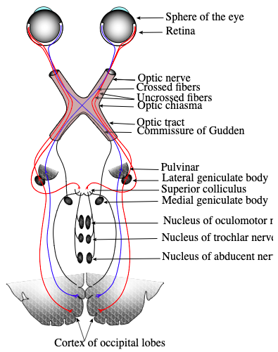
- The nerve then enters the bony optic canal to emerge intracranially on the underside of the front of the brain.
- At this point the optic nerve from each eye comes together and forms an X-shaped structure called the optic chiasm. Here, approximately one-half of the nerve fibres from each eye continue on the same side of the brain, and the remaining nerve fibres cross over at the chiasm to join fibres from the opposite eye on the other side of the brain. This arrangement is essential for producing binocular vision.
- Posterior to the optic chiasm, the nerve fibres travel in optic tracts to various portions of the brain. See image 1.
- Some nerve fibres leave the optic tract without entering the lateral geniculate nuclei and instead enter the brain stem to provide information that ultimately determines pupil size.[3]

Damage[edit | edit source]

Damage to an optic nerve or damage to its pathways to the brain results in loss of vision. At the optic chiasm, each optic nerve splits, and half of its fibers cross over to the other side. Because of this anatomic arrangement, damage along the optic nerve pathway causes specific patterns of vision loss. By understanding the pattern of vision loss, a doctor can often determine where the problem is in the pathway[4].
There are many different types of optic nerve disorders, including:
- Glaucoma is a group of diseases that are the leading cause of blindness in the United States. Glaucoma usually happens when the fluid pressure inside the eyes slowly rises and damages the optic nerve.
- Optic neuritis is an inflammation of the optic nerve. Causes include infections and immune-related illnesses such as multiple sclerosis. Sometimes the cause is unknown.
- Optic nerve atrophy is damage to the optic nerve. Causes include poor blood flow to the eye, disease, trauma, or exposure to toxic substances.
- Optic nerve head drusen are pockets of protein and calcium salts that build up in the optic nerve over time[2][3].

The anatomy of the optic nerve makes it a sensitive marker for problems inside the brain. This nerve connects the back of each eyeball and its retina to the brain. In its short span between the brain and the eye, the optic nerve's whole surface is bathed in cerebral spinal fluid.
- However, even slight increases in the pressure of this fluid, from swelling of the brain, can compress the optic nerve around its whole circumference in a "choking" manner. Some important causes of increased pressure from cerebral spinal fluid and papilledema are brain tumors and brain infections, such as a brain abscess, meningitis or encephalitis. A significant proportion of people who are diagnosed with brain tumors have some evidence of papilledema. A pressure increase resulting from bleeding or from very high blood pressure also can cause papilledema.[5].
- Image 3. Laser Doppler holography of blood flow in the optic nerve head region of the human retina.
References[edit | edit source]
- ↑ Very well health Optic Nerve Available from: https://www.verywellhealth.com/optic-nerve-anatomy-4686150(accessed 4.2.2021)
- ↑ 2.0 2.1 Medline plus Optic Nerve Available from:https://medlineplus.gov/opticnervedisorders.html (accessed 4.2.2021)
- ↑ 3.0 3.1 Britannica Optic Nerve Available from:https://www.britannica.com/science/optic-nerve (accessed 4.2.2021)
- ↑ MSD manauls eye Disorders Available from: https://www.msdmanuals.com/en-au/home/eye-disorders/optic-nerve-disorders/overview-of-optic-nerve-disorders (accessed 4.2.2021)
- ↑ Drugs.com Optic nerve Available from: https://www.drugs.com/health-guide/optic-nerve-swelling-papilledema.html (accessed 4.2.2021)

