Myofibril
Original Editor - Lucinda hampton
Top Contributors - Lucinda hampton and Vidya Acharya
Introduction[edit | edit source]
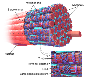
Myofibrils are long contractile fibres, groups of which run parallel to each other on the long axis of the myocytes (long single multinucleated cells that combine to form the muscle). The myocytes run parallel to each other on the long axis of the cell.
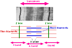
The myofibrils are made up of thick and thin myofilaments, which give the muscle its striped appearance. The thick filaments are composed strands of the protein myosin, and the thin filaments are strands of the protein actin, along with two other muscle regulatory proteins, tropomyosin and troponin. Under the influence of adenosine triphosphate (ATP), actin and myosin form a contractile compound, actomyosin, which is required for muscle contraction.
Sarcomere[edit | edit source]
Myofibrils are essentially polymers, or repeating units, of sarcomeres. The shortening of the individual sarcomeres leads to the contraction of the individual muscle fibres, leading to muscle contractions.[1][2].
- The sarcomere structures give skeletal muscle its striated appearance and are readily visible on electron microscopy.
- The sarcomeric subunits of smooth muscle have no alignment. Hence there are no striations, and hence the cells are called smooth[3].
Myofibrils and Adaption[edit | edit source]
Individual myofibrils (approximately 1 μm in diameter) comprise approximately 80% of the volume of a whole muscle. The number of myofibrils ranges from 50 per myocyte in the muscles of a fetus to approximately 2000 per myocyte in the muscles of an untrained adult. The hypertrophy and atrophy of adult skeletal muscle are associated with certain types of training and disuse and result from the regulation of the number of myofibrils per fiber.
- Growth in the girth of the muscle fibers appears to take place by splitting of the myofibrils which can be stimulated by development of stress on the sarcomere. This adds to the diameter or girth of myofibers without any hyperplasia.
- The growth in length occurs at either end of the fibers and results in addition of new sarcomeres.[4]
References[edit | edit source]
- ↑ Britannica Myofibril Available;https://www.britannica.com/science/myofibril (accessed 9.7.2022)
- ↑ Biology Dictionary Myofibril Available:https://biologydictionary.net/myofibril/ (accessed 9.7.2022)
- ↑ Vedantu Myofibril Available:https://www.vedantu.com/biology/myofibril (accessed 10.7.2022)
- ↑ Pearson AM. Muscle growth and exercise. Critical Reviews in Food Science & Nutrition. 1990 Jan 1;29(3):167-96. Available:https://pubmed.ncbi.nlm.nih.gov/2222798/ (accessed 10.7.2022)


