Lymphoedema
Original Editors - Emily Clark from Bellarmine University's Pathophysiology of Complex Patient Problems project.
Top Contributors - Emily Clark, Lucinda hampton, Admin, Manali K Shah, Fasuba Ayobami, Vidya Acharya, Essam Ahmed, Candace Goh, Kim Jackson, Jonathan Wong, Elaine Lonnemann, Chelsea Mclene, 127.0.0.1 and Wendy Walker
Definition/Description[1][2][edit | edit source]
Lymphatic obstruction is a blockage of a lymph vessel that drains fluid and
immune cells from tissue throughout the body.[3] An obstruction could cause an
impaired contraction of the collecting lymphatics, causing lymphedema which is a
build up of lymph fluid in the soft tissue.[2]
Lymphedema has been classified into grades of severity by the International Society of Lymphology:
- Stage 0 (Latent lymphedema)- Lymph transport capacity is reduced, no clinical edema is present.
- Stage1 - Reversible pitting edema with elevation; Increasing edema with increase in activity, heat, and humidity.
- Stage 2 - Accumulation of protein-rich nonpitting edema with connective scar tissue. Irreversible ; does not resolve overnight; increasingly more difficult to pit.Clinical fibrosis is present.Skin changes present in severe stage 2.
- Stage 3(Lymphostatic Elephantiasis)-Accumulation of protein-rich edema with significant increase in connective and in scar tissue. Severe non-pitting fibrosis edema.Strophic changes (hardening of dermal tissue, skin folds, skin papillomas, and hyperkeratosis) [2][4][1]
Lymphedema can be divided into 2 categories primary/ idiopathic and secondary/ acquired. The primary cause of lymphedema happens due to a malformation of the lymph vessels. Secondary lymphedema is damage that has been done to normal healthy lymph vessels.[2]
Prevalence[edit | edit source]
The incidence of lymphedema is unknown because it goes unreported. When looking at the primary cause 15% of lymphedema cases are reported at birth, 75% during adolescence with a ratio of 4:1 females to males, 10-20% after the age of 35, with 2 % in other syndromes. Secondary causes are just an approximation of the incidences of filariasis, an infection caused by mosquitoes, because it spans across the globe. There was an estimate of 420 million people were exposed to filariasis in Africa in the year 2000 and the WHO estimated 700,000 incidences in the Americas. There are around 3 million cases in the US with 30% of those secondary to breast cancer[2]. One study looked at 300 patients with breast cancer a year later the prevalence of clinically significant lymphedema was 33.5 % and 17.2 % had severe lymphedema. The prevalence of lymphedema was 13.4 % in patients treated with surgery only where as the prevalence was 42.4% in patients treated with surgery and radiotherapy. Post treatment lymphedema continues to be a significant problem following breast cancer therapy. Presence of co-morbid conditions and axillary radiation significantly increases the risk of lymphedema. A combination of axillary dissection and axillary radiation should be avoided whenever feasible to avoid lymphedema. [5]
Characteristics/Clinical Presentation[edit | edit source]
Signs and Symptoms present:
- Swelling in an arm or a leg. It may be the entire limb or only parts . Most likely unilateral, but can be bilateral.
- Heaviness or tightness in involved limb
- Loss of ROM
- Achiness
- Reoccurring infections in the involved limb
- Hardening, thickening, or tightness of the skin[6][2]
- Burning feeling in the legs
- Loss of hair
- Loss of sleep[7]
- Can be fibrosis, pitting edema
- Symptoms can increase during warm weather, menstruation, and if the limb has been left in its depended position.[1]
- If primary and affecting the intestine signs and symptom include; abdominal bloating, diarrhea, and intolerance of fatty foods.[2]
Associated Co-morbidities[edit | edit source]
Risk factors for lymphedema include:
- Removal or radiation of the lymph nodes
- Tumors blocking the flow of lymph fluids
- Overweight or obesity[7]
- Diabetes
- Infection
- Scar tissues of lymph vessels by radiation
- Post surgery inflammation
- Increased Age
- Poor nutrition
- Cancer[8]
In addition, Presence of Lymhedema leads to significant morbidity, activity and participation restriction, reduced quality of life and economic hardship.[9]
Medications[edit | edit source]
There is not a specific lymphedema drug available. Different drugs such as benzopyrones ( Coumarin, Venalot, Daflon, natural ingredients such as rutin, horse chestnut, and rapeseed extract) can affect an increase in proteolysis, which can act to decrease protein concentration and decrease lymphedema. There is a chance for drug toxicity with these drugs. Diuretics that are used for sodium retention edema, are also being prescribed, even though they don’t affect lymphedema. Diuretics can cause an increase risk for electrolyte imbalance. Medications that could cause edema in the legs include NSAIDs, Norvasc for hypertension, Avandia for diabetes, and Lyrica[1] for diabetic neuropathy and shingles. Some chemotherapy medicines may cause a disturbance in behavior that could cause a lack of compliance with treatment.[2]
Diagnostic Test[edit | edit source]
A thorough history must be taken. Palpation of the lymph nodes must be done to see if they are swollen or there are any abnormal changes. Measurements for the swelling limbs should also be taken.[7] In diagnosing the diagnostic tool used is the isotope lymphograph also called lymphoscintigraphy or lymphangioscintigraphy(LAS) is used to determine abnormal lymph nodes and lymphatics. Other imaging tools are MRI, MR Lymphography techniques, computed tomography (CT), Perometry, Bioimpedance analysis, Patient-Reported symptom assessments, ultrasonography(US), and DEXA,etc.[4][9]Progress can be measured by limb circumference and water displacement.[1] Clinicians must be reminded that none of the diagnostic tool is perfect in terms of accuracy.[9]
Etiology/Causes[edit | edit source]
Causes of Lymphedema
Primary Cause
- Unknown
- Hereditary
- Developmental abnormalities:
- Aplasia
- Hypoplasia (75% of cases)
- Hyperplasia(15% of cases)
Secondary Cause
- Filariasis (mosquito bite- parasitic infection)
- Primary or Metastatic Neoplasm (benign or malignant)
- Surgery (lymph node dissection or removal)
- Radiation treatment
- Chemotherapy
- Severe infection
- Other surgeries( multiple abdominal or pelvic surgeries
- Lipedema
- Chronic venous insufficiency
- Liposuction
- Crush injury
- Compound fracture
- Severe Laceration
- Degloving skin injury
- Burns
- Obesity
- Multiparity
- Paralysis
- Prolonged systemic cortisone (cortisone skin)
- HIV/AIDS[2]
The most common cause for secondary lymphedema worldwide is filariasis a parasitic infection caused by mosquitoes.[2] The most common reason for lymphatic obstruction is the removal or enlargement of the lymph nodes in the US.[3]
Systemic Involvement[edit | edit source]
Lymphedema can cause thickening of the dermis and can create ulcerations of the skin. Increased problems healing due to decreased oxygen supply to the tissue. The skin will stretch and cause folds in the skin. It can increase the risk of bacterial or fungal infections underneath the skin folds.
The increased swelling and weight of the limb can create problems in gait, ROM, functions, decreased sensation, balance, strength, increased fatigue and joint contracture due to inactivity.
If primary lymphedema is at birth then it could affect internal organs, including genitals and intestines.
If lymphedema is in the neck, jaw, or shoulders it could involve problems with speech, respiratory function, and swallowing.[2]
Medical Management (current best evidence)[edit | edit source]
- Primary lymphedema can be treated with sclerotherapy to seal a leaky lymph vessels and prevent reflux into the abdomen. Radiation therapy and surgical dissections[2]
- Removal of abnormal lymph vessels[3]
- Microsurgeries performed on lymph vessels to amastomose to a vein or another functional lymph vessel. It has an increased mortality and morbidity rate and are unsuccessful. [2]
Physical Therapy Management (current best evidence)[edit | edit source]
If treatment for cancer is necessary that should be completed first.[2]
Practice pattern H in the Guide to Physical Therapy can help guide your interventions with lymphedema and the complications.[10]
Interventions include:
- Manual lymph drainage (to help improve the flow of lymph from the affected arm or leg from proximal to distal).
- Short/low stretch Compression garment wear following lymphatic drainage.
- Skin Hygiene and care (such as cleaning the skin of the arm or leg daily and moisten with lotion).
- Exercise to improve cardiovascular health and help decrease swelling in some cases.
- Patient education (instruction in proper diet to decrease fluid retention and how to avoid injury and infection, anatomy, and self bandaging).
- Compression pumps
- Psychological and emotional support
- Garment fitting.[8][2][4]
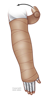
Complex Decongestive Therapy:
- Phase 1:
- Skin care
- Light manual massage (manual lymph drainage)
- ROM
- Compression (multi-layered bandage wrapping, highest level tolerated 20-60 mm Hg)
- Phase 2:

- Compression by low-stretch elastic stocking or sleeve
- Skin care
- Exercise
- Light massage as needed


Contraindications for compression includes arterial disease, painful postphlebitic syndrome, and occult visceral neoplasia.[4]
Differential Diagnosis[edit | edit source]
- Lipedema
- Cellulitis
- Dermatologic Manifestations of Cardiac Disease
- Dermatologic Manifestations of Renal Disease
- Erysipelas
- Filariasis
- Lymphangioma
- Thrombophlebitis
- Venous Insufficiency[11]
Evidence[edit | edit source]
Courneya K, Mackey J, Bell G.Randomized Controlled Trial of Exercise Training in Postmenopausal Breast Cancer Survivors: Cardiopulmonary and Quality of Life Outcomes. Journal of Clinical Oncology, Vol 21, Issue 9 (May), 2003: 1660-1668. http://171.66.121.246/content/21/9/1660.full Accessed on 4/5/2011.
Badger C, Peacock J, Mortimer P. A Randomized, Controlled, Parallel-Group Clinical Trial Comparing Multilayer Bandaging Followed by Hosiery versus Hosiery Alone in the Treatment of Patients with Lymphedema of the Limb. Cancer 2000;88:2832–7.© 2000 American Cancer Society.https://www.cebp.nl/media/m1159.pdf Accessed on 4/5/2011.
McNeely M, Magee D, Lees A, Bagnall K. The Addition of Manual Lymph Drainage to Compression Therapy For Breast Cancer Related Lymphedema: a Randomized Controlled Trial. Volume 86, Number 2, 95-106. http://resources.metapress.com/pdf-preview.axd?code=pm25575l0765836l&size=largest Accessed on 4/5/2011.
Resources
[edit | edit source]
National Cancer Institute http://www.cancer.gov/cancertopics/pdq/supportivecare/lymphedema/Patient/Page2#Section_69
Northwest Medical Center http://northwestmed.com/our-services/lymphedema-management.dot
http://www.vascularweb.org/vascularhealth/Pages/lymphedema.aspx
http://www.medicinenet.com/breast_cancer_and_lymphedema/louisville-ky_city.htm
References[edit | edit source]
- ↑ 1.0 1.1 1.2 1.3 5. The Merck Manuals: The Merck Manual for Healthcare Professionals. Lymphedema. http://www.merck.com/mmpe/sec07/ch081/ch081h.html#sec07-ch081-ch081h-1866. (accessed 5 April 2011)
- ↑ 2.00 2.01 2.02 2.03 2.04 2.05 2.06 2.07 2.08 2.09 2.10 2.11 2.12 2.13 2.14 Goodman C, Fuller K. Pathology: Implications for the Physical Therapist. 3rd ed. St. Louis, MO: Saunders Elsevier: 2009.
- ↑ 3.0 3.1 3.2 PubMed Health.Disease and Conditions: Lymphatic Obstruction. http://www.ncbi.nlm.nih.gov/pubmedhealth/PMH0002106/ (accessed 5 April 2011).
- ↑ 4.0 4.1 4.2 4.3 International Society of Lymphology. Diagnosis and Treatment of Periphreal Lyphology.Lymphology 42 (2009) 51-60. http://www.u.arizona.edu/~witte/ISL.htmDocument. (accessed on 5 April 2011).
- ↑ Deo SV, Ray S, Rath GK, Shukla NK, Kar M, Asthana S, Raina V. Prevalence and Risk Factors for Development of Lymphedema Following Breast Cancer Treatment. Indian J Cancer 2004; 41:8-12. http://www.indianjcancer.com/text.asp?2004/41/1/8/12338 . (accessed 5 April 2011).
- ↑ Mayo Clinic. Lymphedema. http://www.mayoclinic.com/health/lymphedema/DS00609/DSECTION=symptoms (accessed 5 April 2011)
- ↑ 7.0 7.1 7.2 National Cancer Institute: U.S National Institutes of Health. Lymphedema PDQ. http://www.cancer.gov/cancertopics/pdq/supportivecare/lymphedema/Patient/page1. (acceessed 5 April 2011)
- ↑ 8.0 8.1 American Physical Therapy Association. Lymphedema: How Physical Therapist Can Help. http://www.oncologypt.org/mbrs/factsheets/LymphedemaFactSheetFinal.pdf (accessed 5 April 2011).
- ↑ 9.0 9.1 9.2 Levenhagen K, Davies C, Perdomo M, et al. Diagnosis of Upper Quadrant Lymphedema Secondary to Cancer: Clinical Practice Guideline From the Oncology Section of the American Physical Therapy Association. Phys Ther. 2017;97: 729–745.
- ↑ American Physical Therapy Association.Guide to Physical Therapist Practice.2nd ed. Phys Ther 2001. Revised 2003.569-585.
- ↑ Medscape reference. Dermatological Manifestations of Lymphedema Differential Diagnosis. http://emedicine.medscape.com/article/1087313-differential. (accessed 5 April 2011)

