Lumbar Facet Syndrome
Original Editors Kirianne Vander Velden
Top Contributors - Naaike Verhaeghe, Aarti Sareen, Kirianne Vander Velden, Kim Jackson, Rucha Gadgil, Admin, Simisola Ajeyalemi, Wanda van Niekerk, 127.0.0.1, Kai A. Sigel, WikiSysop, Rachael Lowe and Liza De Dobbeleer
Definition/Description[edit | edit source]
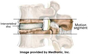
Lumbar facet syndrome means: A dysfunction at the level of the posterior facet joints of the spine. These joints together with the disc form the intervertebral joint. Changes at the level of the posterior facet joints can influence the disc and vice versa.
The term ‘dysfunction’ implies that at a certain level (mostly L4-L5 or L5-S1) these 3 components do not function normally.
In 1976, researchers indicated that the facet joints could be a possible cause for back pain in humans. They injected a intra-articular saline solution which produced severe local and radiating pain in the buttocks and posterior thigh in healthy individuals. Later on studies examined how this pain could be relieved by intra-articular injections, although treatment is nowadays mainly conservative. (31) (32)
The lumbar facet syndrome is a painful irritation of the posterior part of the lumbar spine. Swelling from the surrounding structures, can cause pain due to an irritation of the nerve roots.
Little capsular tears can originate at the level of the posterior facet joints due to a trauma. This can lead to a subluxation of the joint. The synovia that surrounds the joint is damaged and leads to a synovitis. Secondly a hypertonic contraction of the surrounding muscles present itself. This is a protection mechanism that increases the pain. These changes lead to a fibrosis and osteophyte formation.
The most common cause is repetitive micro trauma and as positive result of this chronic degeneration. In daily living this may occur with repetitive extension of the back. So mostly all movements with the arms above the head.
These recurring injuries can happen in sports were it is necessary to make repetitive powerful hyperextensions of the lumbar spine.
An irritation can also occur when the intervertebral disc is damaged and the biomechanics of the joint have changed. In this case the facet joints are exposed to a higher loading.
Clinically Relevant Anatomy[edit | edit source]
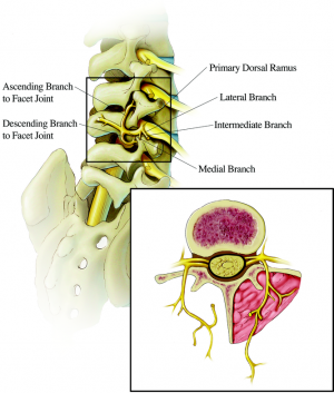
The affected anatomy in this pathology are the facet joints. The lumbar facet joints form the posterolateral articulations. This is by the connection between the vertebral arch of one vertebra to the arch of the adjacent vertebra.They are more properly termed the zygapophyseal joints. It’s a synovial joint. Each facet joint includes a joint space, which is capable of accommodating between 1 and 1.5 ml of fluid, a synovial membrane, hyaline cartilage surfaces, and a fibrous capsule (1mm). This joint is a potential source of pain. Each level of the spine has a three-joint complex to provide for the joint functions. There are two facet joints in the back and a large disc in front. This provides the stability. in the case of lumbar facet joints, the joints get inflamed. Because of the high compressive forces, facet pain in this area is quite common.
Each facet joint acquires a dual innervation from medial branches arising from posterior primary rami. One at the same level, one a level above the z-joint. The medial branches of L1–L4 dorsal rami run across the top of transverse processes one level below the named spinal nerve. After this point each nerve runs downward following the junction of the transverse and superior articular processes. From this point the nerve divides into multiple branches as it crosses the vertebral lamina. The L5 nerve differs itself from the others because it’s the dorsal ramus itself that runs along the junction of the sacral ala and superior articular process of the sacrum. It’s more likely at this level that the dorsal ramus blockades, rather than the medial branch. (9)(48)(49)
Epidemiology /Etiology[edit | edit source]
LBP is a major cause of disability and is the most common reason for medical consultations because this pain problem interferes with activities of daily living and work performance. (43)
LBP is the most common musculoskeletal disorder of industrialized society and the most common cause of disability in persons younger than 45 years, but it can affect people of all ages. (44) Given that 90% of adults experience LBP sometime in their lives, the fact that it is the second leading cause for visits to primary care physicians and the most frequent reason for visits to orthopedic surgeons or neurosurgeons is not surprising. As the primary cause of work-related injuries, LBP is the most costly of all medical diagnoses when time off from work, long-term disability, and medical and legal expenses are taken into account.[1]
Low back pain has a prevalence of 60-70% in industrialized countries. (44)
The lumbosacral facet joint is reported to be the the most common source of mechanical low back pain in 15-45% of patients with chronic LBP. (45) Although, facet joint syndrome is commonly overlooked in patients with chronic low back pain. The authors believe this is because of: (a): the absence of a clinical picture in facet joint syndrome, (b): conventional clinical examination nor radiological examination can diagnosticate facet joint syndrome, (c): only a very small number of doctors perform a manual functional examination to diagnose facet joint syndrome, (d): the diagnostic anasthetic block to confirm the facet joint syndrome is not widely accesible. (46) Ray believed that facet joint–mediated pain is the etiology for most cases of mechanical LBP,[2] whereas other authors have argued that it may contribute to nearly 80% of cases. Thus, the diagnosis and treatment of this entity may help alleviate LBP in a significant number of patients.
According to Nachemson et al, osteoarthritis of the facet joints is irrelevant to diagnosis and can not be used to explain where the patient’s pain is coming from. (47)
Characteristics/Clinical Presentation[1][2][3][edit | edit source]
- [3][4][5] (45)
● Local pressure pain at the level of the affected joint
● Local pressure pain of the M. Multifidi and M. Erector Spinae (when palpating very stiff due to hypertonia)
● Decreased extension and painful restricted locally to the affected joints
● Unilateral abnormal lateroflexion
● Antalgia
● can occur when rising up with a flexed torso
● Sometimes a functional scoliosis in anteflexion
● sensibility/pain local and ipsilateral
● pain in hyperextension
● pain in lateral flexion and y-axis rotation in extension
● pain in hip, bottom and back when lifting a extended leg
● referred pain not further than the knee
● local stiffness
● Kemp’s test positive
● Springing test positive
● Pain: mild – severe, different between patients and within patient.Pain variat during different positions
● Pain on palpation of the facet joints
● By returning from flexion to upright position, the patient climbs up the legs by use of his hands
Increase pain.
- Extension
- Rotation
- Prolonged standing
- Sudden movements
- After rest
- Lateral flexion towards affected side
- Returning from flexed position
- Movements in general
- Sitting, flexion, using a clutch (in a vehicle), coughing and/or sneezing, and walking for a long time
Decrease pain
- Walking
- Lying with knees bent
- Medication
- Supported flexion, sitting, standing with weight on hands and elbows
- Rest
- Lateral bending towards healthy side
- Varying activity
Diagnostic Procedures[edit | edit source]
Lumbar Facet syndrome can not be reliably clinically diagnosed (Jackson RP2 1992). The most used systems to diagnose this syndrome are an X-ray, a computed tomography (CT) scan of the spine or a magnetic resonance imaging (MRI) scan. Plain radiography does not provide information in establishing the diagnosis of the facet joint syndrome. But it may help with the evaluation of the degree of degeneration. Only, once the degeneration is visible on plain radiography, it has already reached an advanced stage. (52)
It’s very important to ask after the following symptoms, this symptoms could include or exclude this diagnosis. If the most of the following symptoms are positive, we can proceed to the next examination.
● The episodes are typically intermittent, and occur a few times per month or year.
● There is a persisting point tenderness overlying the inflamed facet joints and some degree of loss in the spinal muscle flexibility
● There is more discomfort while leaning backward than leaning forward.
● low back pain from the facet joints radiates down into the buttocks and back of upper leg. (with this test we can to the differential diagnosis with discus herniation).
● The pain can also radiate into the shoulders or upper back. (also differential diagnosis with discus herniation). (51)(53)
The working diagnosis of facet pain, based on history and clinical examination, may be confirmed by performing a diagnostic block. This is considered positive when the patient experiences a 50% pain reduction. It involves injecting a medicine into or near the nerves that supply the facet joint. If the pain is not relieved by the injection, it is unlikely that the facet joint is the source of the pain. If these injections help and reduce the pain, we can suggest that the pain comes from the facet joint.Although no single sign or symptom is diagnostic, Jackson et al demonstrated that the combination of the following 7 factors was significantly correlated with pain relief from an intra-articular facet joint injection:
● Older age
● Previous history of LBP
● Normal gait
● Maximal pain with extension from a fully flexed position
● The absence of leg pain
● The absence of muscle spasm
● The absence of exacerbation with a Valsalva maneuver (51)(53)
Differential Diagnosis[edit | edit source]
- ● Lumbosacral Disc Injuries and degeneration[28]
● Lumbosacral Discogenic Pain [22]
● Lumbosacral Radiculopathy
● Lumbosacral Spine Acute Bony Injuries
● Lumbosacral Spine Sprain/Strain Injuries
● Lumbosacral (degenerative) Spondylolisthesis [28]
● Ankylosing spondylitis [22]
● Piriformis Syndrome
● Sacroiliac Joint Injury/Pathology [13][22]
● Inflammatory arthrides (rheumatoid arthritis, psoriatic arthritis, reactive arthritis) [22]
● Spondylarthropathies (ex. osteoarthrosis, synovitis)[22,28]
● Lumbosacral ligamenteous injury[22]
● Myofascial pain[22]
Within the context of facet pathology, inflammatory arthritides, such as rheumatoid arthritis, ankylosing spondylitis, gout, psoriatic arthritis, reactive arthritis, and other spondylarthropathies, as well as osteoarthrosis and synovitis, must also be considered. (50)
Outcome measures[edit | edit source]
NRS pain score ( numeric rating scale): (33F),
Roland disability Questionnaire (54)
Oswestry Disability Questionnaire (54)
Examination[edit | edit source]
Inspection
The inspection include an evaluation of paraspinal muscle fullness or asymmetry, increase or decrease in lumbar lordosis, muscle atrophy, or posture asymmetry.
Patients with chronic facet syndrome may have flattening of the lumbar lordosis and rotation or lateral bending at the sacroiliac joint or thoracolumbar area. (A)
Palpation
The examiner should palpate along the paravertebral regions and directly over the transverse processes because the facet joints are not truly palpable. This is performed in an attempt to localize and reproduce any point tenderness, which is usually present with facet joint–mediated pain.
In some cases, facet joint–mediated pain may radiate to the gluteal or posterior thigh regions. (A)
Range of motion[21,22,25,29,30]
Range of motion should be assessed through flexion, extension, lateral bending, and rotation.
With facet joint–mediated LBP, pain is often increased while standing, with extension, flexion, (axial)rotation of the lumbar spine, and it might be either focal or radiating. Also sitting and rising from sitting can induce the pain. A supine position can improve the pain. Mostly coughing, straightening from flexion, extension combined with extension and hyperextension doesn’t worsen the pain. (A)
Flexibility
Inflexibility of the pelvic musculature can directly impact the mechanics of the lumbosacral spine.
With facet joint pathology, the clinician may find an abnormal pelvic tilt and rotation of the hip secondary to tight hamstrings, hip rotators, and the quadratus, but these findings are nonspecific and can be found in patients with other causes of LBP.
Sensory examination
Sensory examination (ie, light touch and pinprick in a dermatomal distribution) findings are usually normal in persons with facet joint pathology. A Sensory abnormality may indicate an other pathology. [25] (A)
Muscle stretch reflexes
Patients with facet joint–mediated LBP have normal muscle stretch reflexes.[25] Radicular findings are usually absent unless the patient has nerve root impingement from bony overgrowth or a synovial cyst.
Side-to-side asymmetry should lead one to consider possible nerve root impingement. (A)
Muscle strength
Manual muscle testing is important to determine whether weakness is present and whether the distribution of weakness corresponds to a single root, multiple roots, or a peripheral nerve or plexus. Also the examination of functional core strength is important to identify other abnormalities[25]
Muscle strength test results are normal in persons with facet joint pathology[25]; however, subtle weakness of the muscles of the pelvic girdle may contribute to pelvic tilt abnormalities. This subtle weakness may be appreciated with trunk, pelvic, and lower-extremity extension asymmetry. (A)
Straight leg – raise test
While performing the neurological examination by using the Straight leg raise test, patiënts doesn’t feel pain or other sensory abnormalities [25] However, if facet joint hypertrophy or a synovial cyst encroaches on the intervertebral foramen, causing nerve root impingement, this maneuver may elicit a positive response. (A)
Special test for LBA due to facet joint
Kemp’s test positive[6]
Springing test positive[6]
● The pharmacological therapy used by doctors for acute back pains caused by facet joint syndrome is based on administrating muscle relaxants.
● Standard treatment modalities for facet joint syndrome pain include intra-articular steroid injections and radiofrequency denervation of the medial branches innervating the joints. Yet there is much controversy in scientific articles related to this standard treatment.
Cohen S. P. et al. (2007) investigated several publications about the effectiveness of intra-articulair steroid injections and radiofrequency denervation of the medial branches. In uncontrolled studies of people that have never been diagnosed for facet joint syndrome, the long-term relief of back pain after intra-articular steroid injection varies from 18% to 63%.In controlled trials, the results are disputable. In the largest study, the investigators reported no significant difference in outcome between the patients who received large-volume (8 ml) LA and steroids injected into facet joints or around facet joints or intraarticular saline injections. Cohen S. P. et al. (2007) also verified that radiofrequency denervation of the medial branches innervating the joints, is an effective treatment for facet joint syndrome. Unfortunately, there aren’t enough studies that follow the same protocol, to make a conclusion about it.
There is also controversy about the long term effect of radiofrequency denervation. Further research should confirm whether radiofrequency means an effective treatment in people with facet joint syndrome.[8][9][10](53)
Medical Management[edit | edit source]
● RFA (radiofrequency ablation) or RFN (radiofrequency neurotomy) of the medial branches nerves [11,16,18,19,27]
● RFN (radiofrequency neurotomy) followed by microscopic discectomy, again followed by RFN[12,13]
● CRF (continuous radiofrequency thermoagulation): uses a hot probe[15,24]
● Computerized tomography-guided kryorhizotomy: uses a cold probe [17]
● Facet joint injections [20(A),21] Lumbar_Facet_Joint_Injections
● Lumbar Facet Joint Nerve Blocks[26]
Physical Therapy Management[edit | edit source]
When acute signals have disappeared, the underlying cause is treated by physiotherapy:
The first thing you need to do is focusing on education, relative rest, pain relief, maintenance of position that provides comfort, exercise and some modalities. (A)
Education means : to inform your patient. He needs to understand the problems he is having. You may not make him anxious, so a diplomatic approach is necessary to prevent him from catastrophizing. When he is too anxious when he needs to move, you cannot do your exercises. So the kinesiophobia needs to be banned. (A)
Pain relief : Physical therapy includes instruction on proper posture and body mechanics in activities of daily living that protect the injured joints, reduce the symptoms, and prevent further injury.
Maintenance of position that provides comfort : Positions that cause pain (e.g., extension, oblique extension) should be avoided. (G) When your patient is having an antalgic posture, this needs to be treated by giving instructions how he has to keep his back in the right position/straight. He has to keep all physiological curves in his back (cervical lordosis, thoracic kyphosis, lumbar lordosis). These instructions are not only important for passive activities, like sitting and standing, but also for active movements. So when he does a certain movement, he can take a certain posture which will not provoke his symptoms. (A)
Relative rest : Bed rest beyond 2 days is not recommended, activity modification rather than bed rest is strongly recommended. (A)
Now you can start with the exercises.
-low load specific exercises :(J) : the abdominal drawing-in maneuver (ADIM) was introduced to volitionally activate the transversus abdominis (TrA) to correct impairments in motor control. (36)
The purpose of the ADIM is to voluntarily activate TrA thickening and lateral slide while obliquus internus (OI) and externus (OE) should remain relatively unchanged. There is some evidence that ADIM exercises may reduce onset deficits and pain (36).
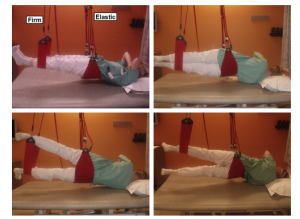
-high load specific exercises : Sling exercises for the lumbopelvic area were performed using the Redcord Trainer. With emphasis on controlling the lumbar spine in a neutral position the subjects performed pain free exercises in closed kinetic chain and under increasing loads. : The overall goal was to improve muscle strength and neuromuscular control. Elastic ropes attached to the band supporting the pelvis were used to ease the load and help subjects maintain a neutral spine position at all times, and for exercises to progress without pain. Exercise progression was achieved by gradually reducing the elasticity of the ropes or increasing the distance (torque) to the distal band.
The overall goal was to improve muscle strength and neuromuscular control. Elastic ropes attached to the band supporting the pelvis were used to ease the load and help subjects maintain a neutral spine position at all times, and for exercises to progress without pain. Exercise progression was achieved by gradually reducing the elasticity of the ropes or increasing the distance (torque) to the distal band. (36)
- trunk, leg and back muscle strengthening exercises. The exercises included sit-ups, push-ups, back rotation, leg press, and pull downs. Number of repetitions/sets and progression of exercises were individually adjusted. (36)
-Therapy for lumbar instability must address not only the lumbar region but also the surrounding anatomical structures such as the muscles of the abdomen and lower extremities Exercises_for_Lumbar_Instability
As therapist you can do passive modalities. You can mobilize the lower back of your patient. In a later stage of the therapy, you can manipulate the lower back.
Reinforcing the muscles of the torso and the pelvis it is also necessary for increase core stability (e.g. by using exercises for pelvis, back and bottom) (A)
Modalities :
-Graded exercises: A graded-exercise intervention emphasizing stabilizing exercises for working patients with nonspecific recurrent LBP seems to improve disability and health parameters such as self-efficacy and physical health, more than do instructions to take daily walks. However, no such positive results emerged for pain over a longer term, or for fear-avoidance beliefs. Although the graded stabilizing exercises seem beneficial in LBP, there is still no clear evidence as to how they affect disability and pain levels.(42)
-Superficial heat and cold : Heat and cold are commonly recommended by clinicians for low back pain. The evidence to support this common practice is not strong. There is moderate evidence that continuous heat therapy reduces pain and disability in the short-term, in a mixed population with acute and subacute low back pain (up to 3 months) and that the addition of exercise to heat wrap therapy further reduces pain and improves function. The application of cold treatment to low back pain is even more limited. No conclusions can be drawn about the use of cold for low back pain. There is conflicting evidence to determine the differences between heat and cold for low back pain. (A) (40)
Therapeutic massage : massage interventions are effective to provide short term improvement of sub-acute and chronic LBP symptoms and decreasing disability at immediate post treatment and short term relief when massage therapy is combined with therapeutic exercise and education (O)
-Balneotherapy : positive effects in reducing pain and improving function , on the patient's’ quality of life, as well as on their analgesic and NSAID requirements. Combined with exercise therapy had advantages than therapy with physical modalities plus exercise in improving quality of life and flexibility of patients with chronic low back pain. (37) (38) (39)
-shockwave (35 ) : Shockwave therapy had shown better long term results compared to facet Joints injections group and little inferior efficacy compared to radiofrequency modulation branch neurotomy. We did not observe any adverse effects and complications in Shockwave therapy group. Moreover, in shockwave therapy and radiofrequency modulation branch neurotomy groups, significant long term improvement in daily activities limitation, was observed Shockwave therapy appears to be a safe and perspective option in the treatment of facet joints pain with negligible side effects.(35)
-spinal manipulation(A) : Spinal_Manipulation
-spinal mobilization(A) : Manual_Therapy_Techniques_For_The_Lumbar_Spine
Current guidlines suggest that non-specific LBP patients should recieve a 12 week course of manual therapy a including spinal manipulation (C)
Spinal manipulative therapy produce slightly better short-term function and perceptions of effect than general exercise, but not better medium or long-term effects, in patients with chronic non-specific back pain (55)
-
Other treatment
-For the treatment of general low-back pain is radiofrequency treatment controversial, for lumbar facet syndrome it seems to have better outcomes. A study that investigated function, pain, and medication use outcomes of radiofrequency ablation for lumbar facet syndrome demonstrated a durable treatment effect of radiofrequency ablation for lumbar facet syndrome at long-term follow-up, as measured by improvement in function, pain, and analgesic use. (33) ( 34)
Key Research[edit | edit source]
Lumbar Facet Syndrome ,Lumbar Facet Syndrome and Therapy,Lumbosacral Facet Syndrome treatment and management , interventional therapies for chronic low back pain,
Heat or cold and low back pain, Low back pain, lumbar facet syndrome and balneotherapy , "Zygapophyseal Joint/abnormalities", "Zygapophyseal Joint/pathology", "Zygapophyseal Joint/physiopathology", Facet Joint Syndrome)
Resources[edit | edit source]
A) http://emedicine.medscape.com/article/94871-treatment
B) Lumbar facet joint injections: https://youtu.be/TcecvNZ9IfM
C) https://www.nice.org.uk/guidance/cg88/resources/commissioning-factsheet-544343293
Clinical Bottom Line[edit | edit source]
Lumbar facet syndrome means: A dysfunction at the level of the posterior facet joints of the spine. These joints together with the disc form the intervertebral joint. Changes at the level of the posterior facet joints can influence the disc and vice versa. The term ‘dysfunction’ implies that at a certain level (mostly L4-L5 or L5-S1) these 3 components do not function normally.
Characteristics are:
● Local pressure pain of the M. Multifidi and M. Erector Spinae
● Local pressure pain at the level of the affected joint
● Decreased extension and painful restricted locally to the affected joints
● Unilateral abnormal lateroflexion/flexed torso
● Antalgia
● sensibility/pain local and ipsilateral
● pain in hyperextension/ lateral flexion and y-axis rotation in extension
● pain in hip, bottom and back when lifting a extended leg
● referred pain not further than the knee
● local stiffness
● Kemp’s test/ springing test positive
● Pain on palpation of the facet joints
● By returning from flexion to upright position, the patient climbs up the legs by use of his hands
Diagnostic procedure: most common they use X-ray and MRI. really important background information is: Older age, previous history of LBP, normal gait, maximal pain with extension from a fully flexed position, the absence of leg pain, the absence of muscle spasm, the absence of exacerbation with a Valsalva maneuver
Examination contains: inspection, palpation, range of motion, flexibility, muscle stretch reflexes, muscle strength, straight leg raise, springing and Kemp’s test.medical management…
When acute signals have disappeared, the underlying cause is treated by physiotherapy:
The first thing you need to do is focusing on education, relative rest, pain relief, maintenance of position that provides comfort, exercise and some modalities.
References[edit | edit source]
- ↑ Prof. Dr. Meeusen R. , Rug- en Nekletsels deel 1: Epidemiologie, anatomie, onderzoek en letsels. Cluwer, Diegem, p 122- 124, 2001
- ↑ Hestbaek L. et Al., The clinical aspects of the acute facet syndrome: results from a structured discussion among European chiropractors. Chiropractic & Osteopathy, 17:2 doi:10.1186/1746-1340-17-2, 2009
- ↑ van Kleef M. et Al., Pain Originating from the Lumbar Facet Joints. Evidence based medicine, World Institute of Pain, Pain Practice, Volume 10, Issue 5,p 459–469, 2010
↑ Hestbaek L. et Al., The clinical aspects of the acute facet syndrome: results from a structured discussion among European chiropractors. Chiropractic & Osteop
5. athy, 17:2 doi:10.1186/1746-1340-17-2, 2009
6. ↑ van Kleef M. et Al., Pain Originating from the Lumbar Facet Joints. Evidence based medicine, World Institute of Pain, Pain Practice, Volume 10, Issue 5,p 459–469, 2010
7. ↑ 6.0 6.1 Hestbaek L. et Al., The clinical aspects of the acute facet syndrome: results from a structured discussion among European chiropractors. Chiropractic & Osteopathy, 17:2 doi:10.1186/1746-1340-17-2, 2009
8. ↑ Mens J.M.. The use of medication in low back pain. Best Pract Res Clin Rheumatol. 2005; 19;609–21.
9. ↑ Cohen S.P., Raja S.N.. Pathogenesis, diagnosis and treatment of lumbar facet joint pain. Anesthesiology. 2007;106;591-614. (Level of evidence 2C)
10. ↑ Lilius G., Laasonen E. M., Myllynen P., Harilainen A., Gronlund G.. Lumbar facet joint syndrome. A randomised clinical trial. Journal of Bone and Joint Surgery. 1989;4;681-684.
11. McCormickZ. et al,Long term funtion, pain and medication use outcomes of radiofrequency ablation for lumbar facet syndrome,J Anesth PMC, 2015 (http://www.ncbi.nlm.nih.gov/pmc/articles/PMC4440581/)
12. Myung Hoon K., et al, Effectiveness of Repeated Radiofrequency Neurotomy for Facet joint Syndrome after Microscopic Discectomy, Korean Journal of spine, 2014 (http://www.ncbi.nlm.nih.gov/pmc/articles/PMC4303287/)
13. Schofferman J. et al, Effectiveness of Repeated Radiofrequency Neurotomy for Lumbar Facet Pain, Spine,2004 (http://ovidsp.tx.ovid.com/sp-3.17.0a/ovidweb.cgi?WebLinkFrameset=1&S=ALLMFPBNDADDBNCPNCJKFDIBIDELAA00&returnUrl=ovidweb.cgi%3f%26Full%2bText%3dL%257cS.sh.22.23%257c0%257c00007632-200411010-00022%26S%3dALLMFPBNDADDBNCPNCJKFDIBIDELAA00&directlink=http%3a%2f%2fgraphics.tx.ovid.com%2fovftpdfs%2fFPDDNCIBFDCPDA00%2ffs047%2fovft%2flive%2fgv024%2f00007632%2f00007632-200411010-00022.pdf&filename=Effectiveness+of+Repeated+Radiofrequency+Neurotomy+for+Lumbar+Facet+Pain.&pdf_key=FPDDNCIBFDCPDA00&pdf_index=/fs047/ovft/live/gv024/00007632/00007632-200411010-00022)
[14]Beresford Z.M. et al., Lumbar facet syndrome, Sports medicine, 2010 (3)
[15]Kroll H., A randomized, double-blind, prospective study comparing the efficacy of continuous versus pulsed radiofrequency in the treatment of lumbar facet syndrome, Journal of clinical Anesthesia, 2008 (2A) http://www.sciencedirect.com.ezproxy.vub.ac.be:2048/science/article/pii/S0952818008002833
[16]Van Wijk R.M.A.W. Radiofrequency Denervation of Lumbar Facet Joints in the Treatment of Chronic Low Back Pain , Journal of pain 2005. (1A) http://www.rmaoem.org/Pdf%20docs/Radiofreq%20denervation%20lumbar%20facet%20rct.pdf
[17] Staender M. et al., Computerized tomography—guided kryorhizotomy in 76 patients with lumbar facet joint syndrome, Journal of neurosurgery: Spine, 2005 http://thejns.org/doi/abs/10.3171/spi.2005.3.6.0444?url_ver=Z39.88-2003&rfr_id=ori:rid:crossref.org&rfr_dat=cr_pub%3dpubmed (3)
[18]Son H.J. The Efficacy of Repeated Radiofrequency Medial Branch Neurotomy for Lumbar Facet Syndrome, Journal of Korean neurosurgical society, 2010(3) http://www.ncbi.nlm.nih.gov/pmc/articles/PMC2966726/
[19]Boswell M. et al., A Systematic Review of Therapeutic Facet Joint Interventions in Chronic Spinal Pain, Pain Physician,2007 (3) http://www.painphysicianjournal.com/current/pdf?article=Nzgw&journal=31
[20] Ribeiro et al.,Effect of Facet Joint Injection Versus Systemic Steroids in Low Back Pain: A Randomized Controlled Trial, Spine, 2013 (1A)
[21]Shulte T.L. Injection therapy of lumbar facet syndrome: a prospective study, Acta Neurochirurgica, 2006
[22] Van Kleef M. et al., Pain Originating from the Lumbar Facet Joints, 0 World Institute of Pain, 2010 (2A)
[23]Cabraja M., et al., The short- and mid-term effect of dynamic interspinous distraction in the treatment of recurrent lumbar facet joint pain, European spine journal, 2009 (2)
[24]Kroll H. et al., A randomized, double-blind, prospective study comparing the efficacy of continuous versus pulsed radiofrequency in the treatment of lumbar facet syndrome, Journal of clinical anesthesia,2008
[25]Beresford Z. et al., Lumbar Facet Syndromes, Current sports medicine reports, 2010
[26]Manchikanti L. et al., Lumbar Facet Joint Nerve Blocks in Managing Chronic Facet Joint Pain: One-Year Follow-up of a Randomized, Double-Blind Controlled Trial, Pain physician journal, 2008 (1A)
[27]Leggett L. et al., Radiofrequency ablation for chronic low back pain: A systematic review of randomized controlled trials, Pain, Research and Management, 2014 (2A)
[28]Boden S. et al., Orientation of the Lumbar Facet Joints: Association with Degenerative Disc Disease, The journal of bone and joint surgery, 1996 (3B)
[29]Revel M. et al., Capacity of the clinical picture to characterize low back pain relieved by facet joint anesthesia: Proposed criteria to identify patients with painful facet joints. Spine 1998; 23 (2C)
[30]Young S. et al., Correlation of clinical examination characteristics with three sources of chronic low back pain, The Spine Journal, 2003 (2C)
(31): Ribeiro, Luiza Helena MD; Furtado, Rita Nely Vilar MD; Konai, Monique Sayuri MD; Andreo, Ana Beatriz MD; Rosenfeld, Andre MD; Natour, Jamil MD, Effect of Facet Joint Injection Versus Systemic Steroids in Low Back Pain: A Randomized Controlled Trial, Volume 38(23), 01 November 2013, p 1995–2002, Level of evidence: 1B
(32): G Lilius; EM Laasonen; P Myllynen; A Harilainen; G Gronlund, Lumbar facet joint syndrome. A randomised clinical trial, J Bone Joint Surg Br August 1989vol. 71-B no. 4 681-684, Level of evidence: 1B
(33) McCormick (ZL, Marshall B, Walker J, McCarthy R, Walega DR. Long-Term Function, Pain and Medication Use Outcomes of Radiofrequency Ablation for LumbarFacetSyndrome. Int J Anesth 2015; 2 (2). PII: 028. (LOE : 2A)
(34). Elias Veizi, MD, PhD; Salim Hayek, MD, PhD, Interventional Therapies for Chronic Low Back Pain, International Neuromodulation Society Neuromodulation 2014; 17: 31–45 (LOE 2A)
(35). Tomas Nedelka 1,2,3, Jiri Nedelka 2, Jakub Schlenker 3, Christopher Hankins 4, Radim Mazanec , Mechano-transduction effect of shockwaves in the treatment of lumbar facet joint pain: Comparative effectiveness evaluation of shockwave therapy, steroid injections and radiofrequency medial branch neurotomy, Neuroendocrinol Lett 2014; 35(5):393–397 (LOE : 2A)
(36) Ottar Vasseljen*, Anne Margrethe Fladmark, Abdominal muscle contraction thickness and function after specific and general exercises: A randomized controlled trial in chronic low back pain patients, Manual Therapy 15 (2010) 482-489 (LOE : 1B)
(37) Nur Kesiktas • Sinem Karakas • Kerem Gun • Nuran Gun • Sadiye Murat • Murat Uludag, Balneotherapy for chronic low back pain: a randomized, controlled study, Rheumatol Int (2012) 32:3193–3199 evidence RCT (LOE : 1B)
(38) Ildiko´ Katalin Tefner • Andra´s Ne´meth • Andrea La´szlo´fi • Tı´mea Kis • Gyula Gyetvai • Tama´s Bender, The effect of spa therapy in chronic low back pain: a randomized controlled, single-blind, follow-up study, Rheumatol Int (2012) 32:3163–3169 (LOE : 1B)
(39) Tefner IK, Németh A, Lászlófi A, Kis T, Gyetvai G, Bender T (2012) The effect of spa therapy in chronic low back pain: a randomized controlled, single-blind, follow-up study. Rheumatol Int 32:3163–3169 (LOE : 1B)
(40) Simon D. French, MPH, BAppSc(Chiro), Melainie Cameron, PhD, BAppSc(Osteo), MHSc(Research), Bruce F. Walker, DC, MPH, DrPH, John W. Reggars, DC, MChiroSc, and Adrian J. Esterman, PhD, AStat, DLSHTM, A Cochrane Review of Superficial Heat or Cold for Low Back Pain, SPINE Volume 31, Number 9, pp 998–1006 (LOE : 1A)
(41). Lucie Brosseau, PhD et al ,Ottawa Panel evidence-based clinical practice guidelines on therapeutic massage for low back pain , Journal of Bodywork & Movement Therapies (2012) 16, 424e455 (LOE :2C)
(42) Eva Rasmussen-Barr, RPT, MSc,*† Bjorn A¨ ng, RPT, PhD,* Inga Arvidsson, RPT, PhD,* and Lena Nilsson-Wikmar, RPT, PhD* , Graded Exercise for Recurrent Low-Back Pain A Randomized, Controlled Trial With 6-, 12-, and 36-Month Follow-ups, SPINE Volume 34, Number 3, pp 221–228 (LOE :1B)
(43): George E. Ehrlich, Low Back Pain, Bulletin of the World Health Organization 2003, 81 (9), pp 671-676, link: http://www.who.int/bulletin/volumes/81/9/Ehrlich.pdf Level of evidence: 1A
(44): Béatrice Duthey, Ph.D, Background Paper 6.24 Low back pain, Priority Medicines for Europe and the World "A Public Health Approach to Innovation", 15 March 2013, pp 3-4, p10. Level of evidence 1A
(45): TM. Markwalder, M. Mérat, The lumbar and lumbosacral facet-syndrome. Diagnostic measures, surgical treatment and results in 119 patients, Acta Neurochirurgica, March 1994, Volume 128, Issue 1, pp 40-46. Level of evidence 2B
(46): Grgić V, Lumbosacral facet syndrome: functional and organic disorders of lumbosacral facet joints, Lijec Vjesn. 2011 Sep-Oct;133(9-10):330-6.) Level of evidence: 1A
(47): GEORGE E. EHRLICH, Back Pain, The Journal of Rheumatology, 2003;30 Suppl 67:26-31. Level of evidence: 1A
(48) Pedersen HE, Blunck CF, Gardner E: The anatomy of lumbosacral posterior rami and meningeal branches of spinal nerves (sinuvertebral nerves). J Bone Joint Surg (Am) 1956; 38:377–91Pedersen, HE Blunck, CF Gardner, E: 3A
(49) Bogduk N: Clinical Anatomy of the Lumbar Spine and Sacrum, 3rd edition. Edinburgh, Churchill Livingstone, 1997, pp 33–42Bogduk, N Edinburgh Churchill Livingstone (level of evidence: 4)
(50) NM Orpen, NC Birch - Journal of spinal disorders & techniques, 2003 - journals.lww.com
(51) Ray C, MD, symptoms and diagnosis of facet joint problems, spine health, 2002.
(52) Jackson, et al,The facet syndrome. Myth or reality?, Clin Orthop Relat Res. 1992 Jun;(279):110-21. (level of evidence: 2C)
(53) Malanga G., Lumbosacral Facet Syndrome Workup, 2015, Medscape. (level of evidence: 2A)
(54) Ronald M., The Roland–Morris Disability Questionnaire and the Oswestry Disability Questionnaire, Spine, 15 December 2000 - Volume 25 - Issue 24 - pp 3115-3124(Loe 5)
(55) Ferreira ML1, Ferreira PH, Latimer J, Herbert RD, Hodges PW, Jennings MD, Maher CG, Refshauge KM ; Comparison of general exercise, motor control exercise and spinal manipulative therapy for chroniclow back pain: A randomized trial.; Pain . 2007 Sep;131(1-2):31-7. (LOE: 1B)


