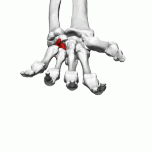
Hamate
Original Editor
Top Contributors - Kim Jackson, Nina Myburg and Patti Cavaleri
Description[edit | edit source]
The hamate bone is one of eight carpal bones that forms part of the wrist joint. The word hamate is derived from the Latin word hamulus which means “a little hook”. It is a wedge-shaped bone with a hook-like process that can be found in the medial side of the wrist.[1] Sometimes it is also called unciform bone.
Structure[edit | edit source]
The hamate has a wedge-like shape with a distinct bony process called hook of hamate that extends from the palmar surface. It is situated in the distal row of carpal bones on the medial side of the wrist. It has multiple articulation surfaces. The hook of hamate can be palpated on the medial side of the wrist inferior and laterally to the pisiform.[1]
Function[edit | edit source]
The hamate is one of the carpal bones that form the carpal arch, wherein the carpal tunnel is situated. It forms part of the ulnar border of the carpal tunnel. It also serves as an attachment for multiple structures such as the transverse carpal ligament (flexor retinaculum). The pisohamate ligament connects the pisiform to the hamate and this forms an osseofibrous tunnel that is called the ulnar canal (Guyon canal). The ulnar nerve passes through this canal between the pisiform and hook of hamate.[1]
Articulations[edit | edit source]
The hamate articulates with the 4th and 5th metacarpals, lunate, triquetrum and capitate. [2]
Muscle attachments[edit | edit source]
- Flexor carpi ulnaris - This is a muscle in the anterior compartment of the forearm. It attaches distally to the pisiform, hook of hamate and 5th metacarpal. It allows wrist flexion and adduction.
- Flexor digiti minimi brevis - This is an intrinsic muscle of the hand. It attaches proximally to the hook of hamate and flexor retinaculum. It allows flexion of the proximal phalanx of the 5th finger.
- Opponens digiti minimi - This is an intrinsic muscle of the hand. It attaches proximally to the hook of hamate and flexor retinaculum. It draws the 5th metacarpal anterior and rotates it to get opposition with the thumb.
See Also[edit | edit source]
References[edit | edit source]
- ↑ 1.0 1.1 1.2 Moore KL, Dalley AF. Clinically Oriented Anatomy. Fifth edition. Philadelphia: Lippincot Williams & Wilkins; 2006
- ↑ Gray H. Anatomy of the Human Body. Twentieth edition. Philadelphia: Lea & Febiger; 1918 Available from: https://www.bartleby.com/107/ [Accessed 30 April 2019]


