Glioblastoma
Original Editors - Simone Potts from Bellarmine University's Pathophysiology of Complex Patient Problems project.
Top Contributors - Simone Potts, Lucinda hampton, Admin, Kim Jackson, Elaine Lonnemann, 127.0.0.1, Wendy Walker and WikiSysop
Definition / Description[edit | edit source]
Glioblastoma Multiforme develops from star-shaped glial cells that support nerve cells. A glioblastoma multiforme is classified as a grade IV astrocytoma. It is also referred to as a glioblastoma or GBM[1]
Glioblastoma multiforme (GBM) is the most common and most malignant of the glial tumors.
- Media attention was brought to this form of brain cancer when Senator Ted Kennedy was diagnosed with glioblastoma and ultimately died from it.
(Photo courtesy of: tedkennedy.us)
Gliomas are a heterogeneous group of neoplasms that differ in location within the central nervous system. There is no particular age or sex distribution. Growth potential, extent of invasiveness, morphological features, tendency for progression, and response to treatments vary between each case diagnosed.
GBM can spread through the brain tissue, but rarely spreads to other areas outside of the central nervous system.
All GBM tumors have abnormal and numerous blood vessels, a common feature of a fast-growing tumor. These blood vessels deliver necessary oxygen and nutrients to the tumors, helping them grow and spread. In addition, these blood vessels easily mix with normal brain tissue and travel away from the main tumor, which makes GBM tumors a challenge to treat.[2]
Prevalence[edit | edit source]
Approximately 60% of the estimated 17,000 primary brain tumors diagnosed in the United States each year are gliomas.
Glioblastoma multiforme is the most frequent primary brain tumor. In most European and North American countries, incidence is approximately 2-3 new cases per 100,000 people per year. [2]
(Photo courtesy of: TextMed)
.
Characteristics / Clinical Presentation[edit | edit source]
• Most invasive type of glial tumor
• Commonly spreads to nearby tissue
• Grows rapidly
• Includes distinct genetic subtypes
• May be composed of many different kinds of cells
• May have evolved from a low-grade astrocytoma or an oligodendroglioma
• Common among men and women in their 50s-70s
• More common in men than women[1]
The most common presentation of patients with glioblastomas is a slowly progressive neurologic deficit, usually motorweakness. However, the most common symptom experienced by patients is headache.
Patients may present with generalized symptoms of increased intracranial pressure (ICP), including headaches, nausea and vomiting, and cognitive impairment.
General symptoms include headaches, nausea and vomiting, personality changes, and slowing of cognitive function.
Headaches can vary in intensity and quality, and they frequently are more severe in the early morning or upon first awakening.
Changes in personality, mood, mental capacity, and concentration can be early indicators or may be the only abnormalities observed.
Focal signs include hemiparesis, sensory loss, visual loss, aphasia, and others.
Seizures are a presenting symptom in approximately 20% of patients with supratentorial brain tumors.[2]
o Increased Intracranial Pressure
o Headache, especially retroorbital; sometimes worse upon awakening, improves during the day
o Vomiting (with or without nausea)
o Visual changes (blurring, blind spots, diplopia, abnormal eye movements)
o Changes in mentation (impaired thinking, difficulty concentrating or reading, memory or speech)
o Personality change, irritability
o Unusual drowsiness, increased sleeping
o Sensory changes
o Muscle weakness or hemiparesis
o Bladder dysfunction
o Increased lower extremity reflexes compared with upper extremity reflexes
o Decreased coordination, gait changes, ataxia
o Positive Babinski reflex
o Clonus (ankle or wrist)
o Vertigo, head tilt [3]
Associated Co-morbidities[edit | edit source]
Some associated co-morbidities include:
- Amnesia
- Blindness
- Cerebellar Ataxia
- Dementia
- Hypertension
- Status epilepticus
- Syncope
- Vomiting (Excess/Chronic)
- Recurrent Meningitis
Click here to see other associated diseases and complications
Medications[edit | edit source]
- Avastin
- Decadron (dexamethazone) - used to reduce swelling around the tumor
- Dilantin (phenytoin) - used to prevent seizures[4]
- Temozolomide - chemotherapy that slows cancer cell growth
- Vincristine - chemotherapy used in conjunction with other chemotherapy drugs
- Patients with GBM could also be prescribed with a number of different pain medicaions.
Diagnostic Tests / Lab Tests / Lab Values[edit | edit source]
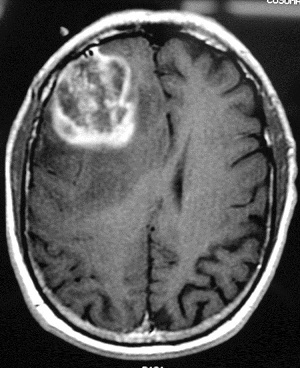
- T1-weighted axial gadolinium-enhanced magnetic resonance image demonstrates an enhancing tumor of the right frontal lobe. Image courtesy of George Jallo, MD. [2]
(Photos courtesy of: Medscape)
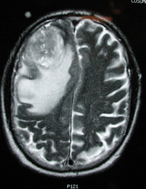
- MRI with or without contrast is the study of choice in diagnosing this disease. These lesions typically have an enhancing ring observed on T1-weighted images and a broad surrounding zone of edema apparent on T2-weighted images.
- Currently, no specific laboratory studies are helpful in making a diagnosis of glioblastoma.
- Positron emission tomography (PET) scans and magnetic resonance (MR) spectroscopy can be helpful in identifying glioblastomas in difficult cases, such as those associated with radiation necrosis or hemorrhage. On PET scans, increased regional glucose metabolism closely correlates with cellularity and reduced survival.[5]
- Patients receiving chemotherapy will present with low white blood cell and platelet counts.
T2 weighted image demonstrates the same lesion as in the previous image, with notable edema and midline shift. This finding is consistent with a high-grade or malignant tumor.
.
Etiology / Causes[edit | edit source]
The etiology of glioblastoma remains unknown in most cases. Familial gliomas account for approximately 5% of malignant gliomas, and less than 1% of gliomas are associated with a known genetic syndrome.
Although concerns have been raised regarding cell phone use as a potential risk factor for development of gliomas, study results have been inconsistent, and this possibility remains controversial. The largest studies have not supported cell phone use as a cancer risk factor.
However, a recently released multinational report concluded that studies that are independent of the telecom industry show that cell phone use may pose a significant risk for brain tumors, and some European countries have taken steps to limit cell phone use by children.[2]
Systemic Involvement[edit | edit source]
Systemic complications include gastritis, pneumonia, sepsis, DVT and pulmonary embolism.
According to Dr. R. Sawaya, in the journal "Neurosurgery," systemic complications occur in approximately eight percent of patients undergoing craniotomy for tumor[6].
Medical Management (current best evidence)[edit | edit source]
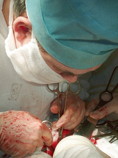
Surgery
- Biopsies
- Partial Resection / Debulking
- Reconstruction
(Photo courtesy of: The Lance Armstrong Foundation)
Radiation
- Traditional Teletherapy - marks made on skin where radiation is delivered
- Brachytherapy - surgically implanted radioactive beads
Chemotherapy
- Can be used in conjunction with radiation
- Patient will become immunodeficient
There are many experimental treatment techniques being developed in hopes to rid patients of GBM tumors. Some of these treatment options are not yet FDA approved. An example of an experimental treatment is: an MRI-guided laser interstitial thermal therapy system called AutoLITT.
(Photo courtesy of: The Internet Journal of Emerging Medical Technologies)
Standard treatment is surgery followed by radiation therapy or a combination of radiation therapy and chemotherapy. If surgery is not an option, the doctor may administer radiation therapy followed by or combined with chemotherapy. Many clinical trials using radiation, chemotherapy, or a combination are available for initial and recurrent GBM. Clinical trials using molecularly targeted therapies showing success in other cancers are also being tested in GBM patients. [1]
Upon initial diagnosis of glioblastoma multiforme (GBM), standard treatment consists of maximal surgical resection, radiotherapy, and chemotherapy with temozolomide.[7]
Physical Therapy Management (current best evidence)[edit | edit source]
No universal restrictions on activity are necessary for patients with glioblastomas. The patient's activity depends on his or her overall neurologic status. The presence of seizures may prevent the patient from driving. In many circumstances, physical therapy and/or rehabilitation are extremely beneficial. Activity is encouraged to reduce the risk of deep venous thrombosis.[7]
A physical therapist may be consulted to assess functional status and provide treatment aimed at maximizing independence and functional capacity. Home or out-patient physical therapy may be recommended to continue to maximize functional mobility. If intensive physical therapy is required, patients may benefit from an inpatient stay at a rehabilitation hospital. Physical therapy evaluation includes identifying what areas may be limiting function: strength, balance, endurance, pain. The physical therapist may prescribe individualized exercises to address the above areas, and may recommend adaptive equipment.[4]
Exercise is good for GBM patients as long as it is to the patient's tolerance. Functional strengthening and aerobic training should be progressed slowly. These patients will benefit greatly from low intensity exercise. Physical therapists should establish the patient's pulmonary function and fitness level (especially if the patient becomes deconditioned after surgery and/or diagnosis).
Patient and family education is crucial to the improvement of each case of GBM. Each patient needs to be made aware of the symptoms, progression and treatment of this cancer.
Differential Diagnosis[edit | edit source]
- Anaplastic astrocytoma
- Cavernous malformation
- Cerebral abscess
- CNS lymphoma
- Encephalitis
- Intracranial hemorrhage
- Metastasis
- Oligodendroglioma
- Radiation necrosis
- Toxoplasmosis[5]
Case Reports / Case Studies[edit | edit source]
- Glioblastoma Multiforme: A Case Study [view case study in The Internet Journal of Advanced Nursing Practice]
- Case Study: Glioblastoma Multiforme [view case study at Medicor Cancer Centres]
- Well-Circumscribed, Minimally Enhancing Glioblastoma Multiforme of the Trigone: A Case Report and Review of the Literature [view article in the American Journal of Neuroradiology]
- Case-Control Study of Use of NSAIDs and GBM [view case study in the American Journal of Epidemiology]
Resources
[edit | edit source]
- Proceedings of the National Academy of Sciences: http://www.ncbi.nlm.nih.gov/pmc/articles/PMC33993/
- American Brain Tumor Association: http://www.abta.org
- American Cancer Society, Inc.: http://www.cancer.org
- National Cancer Institute: http://www.cancer.gov
- National Brain Tumor Society: http://www.braintumor.org
- MGH Brain Tumor Center: http://brain.mgh.harvard.edu/ [(617) 724-8770]
Recent Related Research (from Pubmed)[edit | edit source]
Failed to load RSS feed from http://eutils.ncbi.nlm.nih.gov/entrez/eutils/erss.cgi?rss_guid=1zepmoDGp3XTKZAQU0EPaK8ExMkl8HQAfYYAnfMDQ66fj3MQIM: Error parsing XML for RSS
References[edit | edit source]
- ↑ 1.0 1.1 1.2 Glioblastoma Multiforme (GBM). National Brain Tumor Society. Available at http://www.braintumor.org/patients-family-friends/about-brain-tumors/tumor-types/glioblastoma-multiforme.html?gclid=CNGRyOuN2KcCFSVe7AodwCdk8Q. Accessed March 29, 2011.
- ↑ 2.0 2.1 2.2 2.3 2.4 Glioblastoma Multiforme (background). Medscape. Available at http://emedicine.medscape.com/article/283252-overview. Accessed March 29, 2011.
- ↑ Goodman C, Snyder T. Differential Diagnosis for Physical Therapists: Screening for Referral. St. Louis, MO: Saunders Elsevier: 2007.
- ↑ 4.0 4.1 Glioblastoma multiforme and anaplastic gliomas: A patient guide. Massachusetts General Hospital (Brain Tumor Center). Available at http://brain.mgh.harvard.edu/patientguide.htm. Accessed March 31, 2011.
- ↑ 5.0 5.1 Glioblastoma Multiforme (background). Medscape. Available at http://emedicine.medscape.com/article/283252-diagnosis. Accessed March 29, 2011.
- ↑ Glioblastoma Multiforme Surgery Complications. Livestrong.com. Available at http://www.livestrong.com/article/190510-glioblastoma-multiforme-surgery-complications/. Accessed April 1, 2011.
- ↑ 7.0 7.1 Glioblastoma Multiforme Treatment & Management (medical care). Medscape. Available at http://emedicine.medscape.com/article/283252-treatment. Accessed April 3, 2011.


