Glial Cells
Original Editor - Lucinda hampton
Top Contributors - Lucinda hampton
Introduction[edit | edit source]
The brain is made up of more than just neurones. Although there are about 86-100 billion neurons in the brain, there are about the same number of glial cells in the brain.
- Glial cells, or neuroglia, are cells that surround the neurones of the central nervous system embedded between them, providing both structural and physiological support[1]
- Although glia cells do not carry nerve impulses (action potentials) they do have many important functions. In fact, without glia, the neurones would not work properly[2].
- The term glia is from the Ancient Greek for "glue", as initially these cells were thought to merely act as supporting structures for neurones.
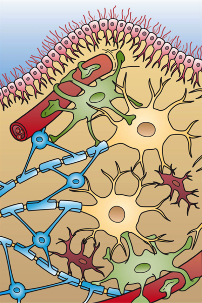
Image: Shows the four different types of Glial cells found in the central nervous system: Ependymal cells (light pink), Astrocytes (green), Microglial cells (red), and Oligodendrocytes (light blue).
Each Glial cell type has specific distribution and function. are broadly divided into:
1. Macroglia [Astrocytes; Oligodendrocytes; Ependymal cells (ependymocytes, tanycytes, choroidal epithelial cells)]
2. Microglia.
Interesting Facts[edit | edit source]
- Glial cells are fascinating and important due to their structural diversity, functional versatility and their ability to change the behavior of firing neurones even though they cannot discharge electrical impulses of their own.
- The have a glymphatic pathway that functionally represents the brain’s lymphatic system[3],
- Glial cells guide early brain development and keep their fellow brain cells healthy throughout life.
- Glia are not mere structural filler, but (as the origin of their name implies, Greek for glue) they help keep things together
- Scientists have shown that glia are, functionally, the brain's other half[4].
Astrocytes[edit | edit source]
Astrocytes are a subtype of glial cells that make up the majority of cells in the human central nervous system (CNS).
- They are star-shaped cells that provide physical and nutritional support for neurons: clean up brain "debris"; transport nutrients to neurons; hold neurons in place; digest parts of dead neurons; regulate content of extracellular space[2]. Unlike neurons and other cells in the nervous system, they do not conduct electrical signals. Instead, they ensure the continued function of their signal-relaying counterparts.
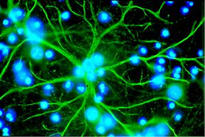
- Image R: Astrocyte in culture emitting many stellate extensions (in green). The blue color represents the nuclei of other cells present in the culture.
The above functions include performing metabolic, structural, homeostatic, and neuroprotective tasks such as:
- Clearing excess neurotransmitters,
- Stabilizing and regulating the blood-brain barrier (they are highly branched and contribute to the blood-brain barrier, the subpial glia limitans,)
- Promoting synapse formation.
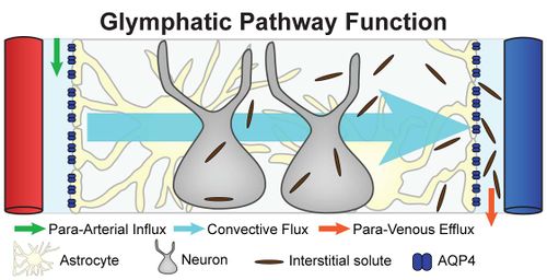
- Regulate extracellular fluid transport known as the glymphatic pathway. The glymphatic pathway has only recently been described and functionally represents the brain’s lymphatic system, although no anatomical structure equivalent to the peripheral lymphatic system is present within the brain parenchyma.[3]
- Metabolic exchange between neurones and capillaries
- Responding to mechanical and biochemical insults to the brain (e.g. gliosis)
The broad and extensive involvement of astrocytes in many aspects of CNS development and function suggests that they carry implications in an array of different nervous system disorders.
- Any condition or stimulus that would impair the ability of astrocytes to function would effectively disturb the blood-brain barrier, synapse formation and stabilization, signaling efficiency, energy metabolism, and fluid-ion homeostasis in the CNS.
- Deficiencies in these functions could potentially cause serious damage to the CNS[5].
Oligodendrocytes[edit | edit source]
Provide the insulation (myelin) to neurones in the central nervous system[2]. Image R: Rat oligodendrocyte development, IF staining for Olig2 and CNPase
- Oligodendrocytes and Schwann cells are the myelin-forming cells of the nervous system. Schwann cells are present in the periphery, and oligodendrocytes are in the central nervous system.
- Myelin is composed of layered phospholipid membranes and serves to support and insulate axons, allowing for faster impulse transduction. Saltatory conduction occurs as the impulses jump across sodium ion-rich nodes of Ranvier. One oligodendrocyte myelinates multiple axons.
- Oligodendrocytes are incapable of replication upon injury[6].
Ependymal Cells[edit | edit source]
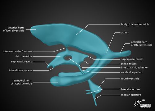
Ependymal cells, which create cerebral spinal fluid (CSF), line the ventricles of the brain and central canal of the spinal cord. These cells are cuboidal to columnar and have cilia and microvilli on their surfaces to circulate and absorb CSF.
Image R: Diagram of the anatomy of the ventricular system of the brain
Ependymal cells encompass three types of cells:
- Ependymocytes: line the ventricles of the brain and central canal of the spinal cord. The are relatively abundant and are involved in the connection between the CSF and nervous tissue
- Tanycytes: line the floor of the third ventricle overlying the median eminence of the hypothalamus
- Choroidal epithelial cells: line the surface of the choroid plexus and are involved in the regulation of cerebrospinal fluid (CSF) contents[6].
Microglia[edit | edit source]
Microglia are the mesoderm-derived, resident macrophages of the CNS. As such, they phagocytose and remove foreign or damaged material, cells, or organisms.[6] They are small, relatively sparse cells.
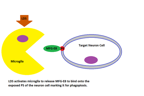
- Microglia act as the brain's resident cleanup squad by phagocytosing apoptotic cells, plaques, and pathogens. Because they can prune and reshape synapses, microglia may also be influential in the pathogenesis of psychiatric illnesses[7]
- Microglia have specific receptors on their surface which recognise distress signals from other cells. These signals attract microglia to the site of the problem. When the brain’s balance is disturbed (usually as a result of inflammation), living neurons can become stressed and produce these signals. This may cause them to be eaten alive by microglia. As neurons are killed, the connections they have with other neurons are also eliminated, which can cause severe issues in brain connectivity and functions.
- With chronic stress, ageing and neurodegenerative disorders microglia may become more aggressive and less easy to regulate which makes them dangerous for the brain. In these cases, microglia can increase in numbers, unnecessarily kill nearby cells, and may contribute to making the brain even more inflamed by secreting inflammatory molecules[8].
- In vivo imaging studies have revealed that in the resting healthy brain, microglia are highly dynamic, moving constantly to actively survey the brain parenchyma.
- Microglia seem to be particularly involved in monitoring the integrity of synaptic function, optimizing different brain circuits to enable cognitive development (microglia cells eliminate previously-formed synapses that are no longer useful).[9]
Glymphatic system[edit | edit source]
The glymphatic system is a recently discovered macroscopic waste clearance system that utilizes a unique system of perivascular channels, formed by astroglial cells, to promote efficient elimination of soluble proteins and metabolites from the central nervous system. Besides waste elimination, the glymphatic system may also function to help distribute non-waste compounds, such as glucose, lipids, amino acids, and neurotransmitters related to volume transmission, in the brain.
The glymphatic system function mainly during sleep and is largely disengaged during wakefulness. The biological need for sleep across all species may therefore reflect that the brain must enter a state of activity that enables elimination of potentially neurotoxic waste products, including β-amyloid.
The glymphatic function is suppressed in various diseases this failure of glymphatic function in turn might contribute to pathology in neurodegenerative disorders, traumatic brain injury and stroke[10].
Image: A schematic illustrating the anatomical components of the glymphatic clearance pathway.
- Aquaporin-4 (AQP4) is one of the most abundant molecules in the brain and is particularly prevalent in astrocytic membranes at the blood-brain and brain-liquor interfaces. While AQP4 has been implicated in a number of pathophysiological processes, its role in brain physiology remains elusive. Evidence suggests that AQP4 is involved in such diverse functions as regulation of extracellular space volume, potassium buffering, cerebrospinal fluid circulation, interstitial fluid resorption, waste clearance, neuroinflammation, osmosensation, cell migration, and Ca(2+) signaling. AQP4 is also required for normal function of the retina, inner ear, and olfactory system[11].
Radial Glia[edit | edit source]
Definition: Glial cells that are involved in neurogenesis. Radial glia also act as scaffolds along which new neurons can travel from their site of origin to their final destination in the brain.
- Responsible for producing all of the neurons in the cerebral cortex.
- Pivotal cell type in the developing central nervous system (CNS) involved in key developmental processes, from patterning and neuronal migration to their recently discovered role as precursors during neurogenesis
- Believed to be a type of stem cell, meaning that they create other cells. In the developing brain, they're the "parents" of neurons, astrocytes, and oligodendrocytes.
- Their role as stem cells, especially as creators of neurons, makes them the focus of research on how to repair brain damage from illness or injury. Later in life, they play roles in neuroplasticity as well[12][13].
References[edit | edit source]
- ↑ Radiopedia Glial cells Available from: https://radiopaedia.org/articles/glial-cells(last accessed 18.12.2020)
- ↑ 2.0 2.1 2.2 Washington Faculty neuroscience for kids Available from:https://faculty.washington.edu/chudler/glia.html (accessed 18.12.2020)
- ↑ 3.0 3.1 radiopedia glymphatic pathway Available from:https://radiopaedia.org/articles/glymphatic-pathway?lang=gb (accessed 18.12.2020)
- ↑ Yuhas D, Jabr F. Know Your Neurons: What Is the Ratio of Glia to Neurons in the Brain?.Available from; https://blogs.scientificamerican.com/brainwaves/know-your-neurons-what-is-the-ratio-of-glia-to-neurons-in-the-brain/(last accessed 18.12.2020)
- ↑ Wei DC, Morrison EH. Histology, Astrocytes. StatPearls [Internet]. 2020 Jun 30.Available from: https://www.ncbi.nlm.nih.gov/books/NBK545142/(accessed 18.12.2020)
- ↑ 6.0 6.1 6.2 Ludwig PE, Das JM. Histology, Glial Cells. StatPearls [Internet]. 2020 Jun 19.Available from:https://www.ncbi.nlm.nih.gov/books/NBK441945/ (accessed 18.12.2020)
- ↑ Scanlon ST. Depressing effects of microglia. Science. 2020 Apr 17;368(6488):279-80.Available from;https://science.sciencemag.org/content/368/6488/279.2.full (last accessed 18.12.2020)
- ↑ The Conversation Microglia: the brain’s ‘immune cells’ protect against diseases – but they can also cause them. Available from:https://theconversation.com/microglia-the-brains-immune-cells-protect-against-diseases-but-they-can-also-cause-them-139232 (last accessed 18.12.2020)
- ↑ Wake H, Moorhouse AJ, Nabekura J. Functions of microglia in the central nervous system-beyond the immune response. Neuron glia biology. 2011 Feb 1;7(1):47.Available from:https://www.cambridge.org/core/journals/neuron-glia-biology/article/abs/functions-of-microglia-in-the-central-nervous-system-beyond-the-immune-response/B262044DDADE74974C8A268DD258B29A (accessed 18.12.2020)
- ↑ Jessen NA, Munk AS, Lundgaard I, Nedergaard M. The glymphatic system: a beginner’s guide. Neurochemical research. 2015 Dec;40(12):2583-99.Available from:https://www.ncbi.nlm.nih.gov/pmc/articles/PMC4636982/ (accessed19.12.2020)
- ↑ Nagelhus EA, Ottersen OP. Physiological roles of aquaporin-4 in brain. Physiological reviews. 2013 Oct;93(4):1543-62.Available from:https://pubmed.ncbi.nlm.nih.gov/24137016/ (accessed 19.12.2020)
- ↑ Very Well Health What are glial cells and what do they do Available from:https://www.verywellhealth.com/what-are-glial-cells-and-what-do-they-do-4159734 (accessed 19.12.2020)
- ↑ Psychology Wiki. Radial Glia Available from: https://psychology.wikia.org/wiki/Radial_glial_cells (accessed 19.12.20200
