Fracture Complications
Original Editor - lucinda hampton
Top Contributors - Lucinda hampton, Naomi O'Reilly and Ewa Jaraczewska
Introduction[edit | edit source]
Most bone injuries heal normally. But some patients do experience complications during the healing process.
Complications of fractures fall into two categories: early and delayed.
- Early complications include shock, fat embolism, compartment syndrome, deep vein thrombosis, thromboembolism (pulmonary embolism), disseminated intravascular coagulopathy, and infection.
- Delayed complications include delayed union and nonunion, avascular necrosis of bone, reaction to internal fixation devices, complex regional pain syndrome, and heterotrophic ossification.[1]
Early Complications[edit | edit source]
Early complications include shock, fat embolism, compartment syndrome, deep vein thrombosis, thromboembolism (pulmonary embolism), disseminated intravascular coagulopathy, and infection.
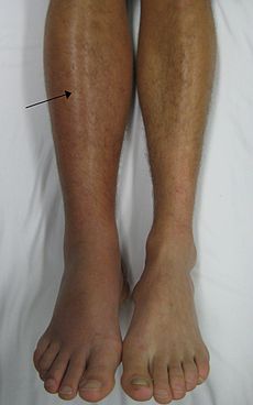
Image 2: DVT in the right leg with swelling and redness
- Shock: Hypovolemic or traumatic shock resulting from hemorrhage and from loss of extracellular fluid into damaged tissues may occur in fractures of the extremities, thorax, pelvis, or spine. Because the bone is very vascular, large quantities of blood may be lost as a result of trauma, especially in fractures of the femur and pelvis. Treatment of shock consists of restoring blood volume and circulation, relieving the patient’s pain, providing adequate splinting, and protecting the patient from further injury and other complications.
- Fat Embolism Syndrome: After fracture of long bones or pelvis, multiple fractures, or crush injuries, fat emboli may develop. Fat embolism syndrome occurs most frequently in young adults (typically those 20 to 30 years of age) and elderly adults who experience fractures of the proximal femur. At the time of fracture, fat globules may move into the blood because the marrow pressure is greater than the capillary pressure or because catecholamines elevated by the patient’s stress reaction mobilize fatty acids and promote the development of fat globules in the bloodstream. The fat globules (emboli) occlude the small blood vessels that supply the lungs, brain, kidneys, and other organs. The onset of symptoms is rapid, usually occurring within 24 to 72 hours, but may occur up to a week after injury. Presenting features include hypoxia, tachypnea, tachycardia, and pyrexia.
- Deep vein thrombosis (DVT), thromboembolism, and pulmonary embolus (PE) are associated with reduced skeletal muscle contractions and bed rest. Patients with fractures of the lower ex-tremities and pelvis are at high risk for thromboembolism. Pulmonary emboli may cause death several days to weeks after injury.
- Disseminated intravascular coagulopathy (DIC) includes a group of bleeding disorders with diverse causes, including massive tissue trauma. Manifestations of DIC include ecchymoses, unexpected bleeding after surgery, and bleeding from the mucous membranes, venipuncture sites, and gastrointestinal and urinary tracts.
- Infection: All open fractures are considered contaminated. Surgical internal fixation of fractures carries a risk for infection. Infections must be treated promptly. Antibiotic therapy must be appropriate and adequate for prevention and treatment of infection[1].
Delayed Complications[edit | edit source]
Delayed complications include delayed union and nonunion, avascular necrosis of bone, reaction to internal fixation devices, complex regional pain syndrome, and heterotrophic ossification.
- Delayed union occurs when healing does not occur at a normal rate for the location and type of fracture. Delayed union may be associated with distraction (pulling apart) of bone fragments, systemic or local infection, poor nutrition, or comorbidity (eg, diabetes mellitus; autoimmune disease). Eventually, the fracture heals.
- Nonunion results from failure of the ends of a fractured bone to unite. The patient complains of persistent discomfort and ab-normal movement at the fracture site. Factors contributing to union problems include infection at the fracture site, interposition of tissue between the bone ends, inadequate immobilization or manipulation that disrupts callus formation, excessive space between bone fragments (bone gap), limited bone contact, and impaired blood supply resulting in avascular necrosis.
- Avascular necrosis occurs when the bone loses its blood supply and dies. It may occur after a fracture with disruption of the blood supply (especially of the femoral neck). The patient develops pain and experiences limited movement. X-rays reveal calcium loss and structural collapse. Treatment generally consists of attempts to revitalize the bone with bone grafts, prosthetic replacement, or arthrodesis (joint fusion).
- Reaction to Internal fixation device: Internal fixation devices may be removed after bony union has taken place. In most patients, however, the device is not removed unless it produces symptoms. Pain and decreased function are the prime indications that a problem has developed[1].
Signs and Symptoms[edit | edit source]
It’s important to know the warning signs of a bone healing complication. Receiving prompt care is critical to treating complications. S &S include:
- Chronic pain
- Drainage from a wound
- Fever
- Swelling
- Limping[2]
Patient-Related Risk Factors[edit | edit source]
Certain patient-related characteristics influence the development of fracture-healing complications in general, even though specific healing complications may differ by their mechanism.
- Diabetes, NSAID use, and a recent motor vehicle accident are most consistently associated with an increased risk of a fracture-healing complication, regardless of fracture site or specific fracture-healing complication. [3]
- In delayed union and non-union identified risk factors include: age; lower limb > upper limb; open fractures; infection; diabetes; smoking; poor blood supply[4].
- Fractures in obese children have a higher rate of complications independently from conservative or surgical treatment. Surgical indications are more common than in normal weighted children and are generally more invasive. [5]
References[edit | edit source]
- ↑ 1.0 1.1 1.2 Brunner LS, Smeltzer SC, Suddarth DS. Brunner & Suddarth's textbook of medical-surgical nursing; Vol. 1. Language. 2010;27:1114-2240p. Available:https://www.brainkart.com/article/Fracture-Healing-and-Complications--Early-and-Delayed-_32596/ (accessed 27.10.2021)
- ↑ Henry Ford Health Systems Bone Healing Complications Available:https://www.henryford.com/services/orthopedics/broken-bones-trauma/complications-healing-bones (accessed 27.10.2021)
- ↑ Hernandez RK, Do TP, Critchlow CW, Dent RE, Jick SS. Patient-related risk factors for fracture-healing complications in the United Kingdom General Practice Research Database. Acta orthopaedica. 2012 Dec 1;83(6):653-60. Available:https://pubmed.ncbi.nlm.nih.gov/23140093/ (accessed 27.10.2021)
- ↑ Coughlin T. Initial Management of Trauma.Available: http://www.learnorthopaedics.com/Learn_Orthopaedics/Musculoskeletal_Trauma_files/Fracture%20Complications.pdf(accessed 27.10.2021)
- ↑ Donati F, Costici PF, De Salvatore S, Burrofato A, Micciulli E, Maiese A, Santoro P, La Russa R. A perspective on management of limb fractures in obese children: is it time for dedicated guidelines?. Frontiers in pediatrics. 2020 May 8;8:207. Available:https://www.frontiersin.org/articles/10.3389/fped.2020.00207/full (accessed 28.10.2021)


