External Carotid Artery
Original Editors - Asma Alshehri
Top Contributors - Asma Alshehri, Lucinda hampton, Kim Jackson, Evan Thomas and Khloud Shreif
Description[edit | edit source]
The external carotid artery is one of the two terminal branches of the common carotid artery which is smaller than the other branch (the internal carotid artery).[1]
Course[edit | edit source]
At the location of the upper border of the thyroid cartilage (typically at the level of the fourth or fifth cervical vertebra), the common carotid arteries bifurcate into the ECA and ICA.
This bifurcation point is clinically significant as it serves as a point for the location of the "carotid body," a chemoreceptor, and the "carotid sinus," a baroreceptor. The carotid body chemoreceptor is sensitive to decreased PO2, increased PCO2, and decreased pH of blood, and is responsible for alerting the brain to change the respiratory rate. The carotid sinus baroreceptors respond to changes in the stretch of the blood vessel and are responsible for detecting changes and maintaining blood pressure.
After its division, the ECA exits the sheath to provide oxygenated blood to the face and neck (the ICA continues in the carotid sheath to enter the carotid canal within the temporal bone).[1][2]
Branches and Divisions[edit | edit source]
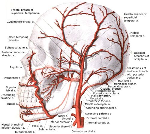
The ECA has eight branches, which anastomose with the branches from the contralateral external carotid, allowing for collateral circulation: These branches include
- Superior thyroid artery
- Ascending pharyngeal artery
- Lingual artery
- Facial artery
- Occipital artery
- Posterior auricular artery
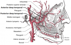
- Maxillary artery
- Superficial temporal artery[1][2]
It branches are also involved in Temporal Arteritis (Giant Cell Arteritis)
Supply[edit | edit source]
It gives supply to the neck, face, and base of the skull.
Clinical Relevance[edit | edit source]
Understanding the vascular supply of the carotid arteries is important. Branches of the ECA supply
- Muscles of mastication receive vascular supply from the maxillary artery and facial artery
- Facial artery also supplies muscles of facial expression.
- Occipital artery supplies blood to the sternocleidomastoid, trapezius, and deep muscles of the back.
- Superficial temporal artery supplies the temporal muscle.
References[edit | edit source]
- ↑ 1.0 1.1 1.2 External carotid artery, https://radiopaedia.org/articles/external-carotid-artery-1 (accessed 30 may 2017)
- ↑ 2.0 2.1 Sethi D, Gofur EM, Waheed A. Anatomy, Head and Neck, Carotid Arteries. InStatPearls [Internet] 2019 Jul 22. StatPearls Publishing. Available from:https://www.ncbi.nlm.nih.gov/books/NBK545238/ (last accessed 27.5.2020)
- ↑ AnatomyZone. External Carotid Branches - 3D Anatomy Tutorial. Available from: http://www.youtube.com/watch?v=K-qtoLS3L4w[last accessed 27/5/2020]


