Extensor Tendon Injuries of the Hand
Original Editors - Sofie Christiaens as as part of the Vrije Universiteit Brussel's Evidence-based Practice project
Top Contributors - Wanda van Niekerk, Sofie Christiaens, Kim Jackson, Laura Ritchie, Andeela Hafeez, Admin, WikiSysop, Shaimaa Eldib, Tarina van der Stockt, Claire Knott, Jess Bell, Anas Mohamed, 127.0.0.1, Evan Thomas, Mariam Hashem and Simisola Ajeyalemi
Definition/Description[edit | edit source]
An extensor tendon injury is a cut or tear to one of the extensor tendons. Due to this injury, there is an inability to fully and forcefully extend the wrist and/or fingers.
Clinically Relevant Anatomy[edit | edit source]
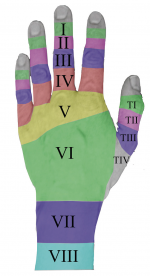
Extensor tendons are located at the dorsal region of the hand and fingers. The function of these tendons is to extend the wrist and the fingers. According to Kleinert and Verdan (1983), there are eight anatomic zones in which the extensor mechanism is divided[1][2] ;
Zone I: DIP joint
Zone II: middle phalanx
Zone III: PIP joint
Zone IV: proximal phalanx
Zone V: MCP joint
Zone VI: metacarpals
Zone VII: wrist (carpus and extensor retinaculum)
Zone VIII: distal third of the forearm[3]
Epidemiology /Etiology[edit | edit source]
The extensor tendons of the hand are located superficially, so they are very susceptible to injuries. An-other reason is the lack of subcutaneous tissue between the tendons and the overlying skin[4]. Possible mechanisms are sharp object direct lacerations, burns, blunt trauma, bites, crush injuries, avulsions and deep abrasions[5]. Closed injuries arise usually under situations of extreme load[2]. This results in ripping the tendons apart from their attachment of the bone.
Characteristics/Clinical Presentation[edit | edit source]
Dependent on the zone of injury, different characteristics are shown.
- Zone I: Mallet finger
- Zone II: no complete rupture of the tendon, but partially injured[2]
- Zone III: Disruption of the central slip, also called a Boutonnière deformity or jammed finger. This is characterised by a flexed position of the PIP joint and an extension or hyperextension of the DIP joint[6].
- Zone IV: injuries are frequently partial, with or without loss of extension at the PIP joint[3]
- Zone V: fight bite injuries (open injuries) or non-fight bite injuries (e.g. blunt trauma): a possible effect of such an injury is a rupture of the sagittal bands, attended with following extensor tendon subluxation[3]. This is presented as a difficulty to actively straighten the flexed MCP joint[2].
- Zone VI: the MCP joint can still be extended via the juncturae tendinum.
- Zone VII: physical injury to the extensor retinaculum[3]
Differential Diagnosis[edit | edit source]
- Mallet Finger refers to a drooping end-joint of a finger. This happens when an extensor tendon has been cut or torn from the bone (Figure 2). It is common when a ball or other object strikes the tip of the finger or thumb and forcibly bends it.
- Boutonnière Deformity describes the bent-down (flexed) position of the middle joint of the finger. Boutonniere can happen from a cut or tear of the extensor tendon .[7]
- Cuts on the back of the hand can injure the extensor tendons. This can make it difficult to straighten your fingers[7]
- Trigger finger (no passive movement possible)
- PIN syndrome: tenodesis effect present - not present with rupture[8]
Diagnostic Procedures[edit | edit source]
Radiographs are recommended because associated injuries of surrounding structures are common. For example, it can be that a piece of bone is pulled off with the tendon[3]. The rupture of the tendon isn’t visible at a radiograph because it’s a soft tissue.
Outcome Measures[edit | edit source]
- Disability of Arm, Shoulder, and Hand questionnaire (DASH)
- Quick DASH –This outcome measure is a shortened version of the DASH and is used to determine the patient’s physical function and symptoms.
- Gartland and Werley Score – This is one of the most widely used outcome measures used in the clinic to evaluate wrist and hand function.
Examination[edit | edit source]
Examination of extensor tendon injuries contains different points of interest. First, the wound characteristics should be evaluated e.g.such as size and location to give the physical therapist has an idea of which structures may have been damaged. Next, the function of the fingers and wrist will be tested in three ways: passively, actively and then with resistance. It is important that each finger is tested separately because the juncturae tendinum between the communis tendons can mask a dysfunction. Furthermore, complete neurovascular examinations should be done.
Specific for zone III injuries, the Elson test can be used[3].
| [9] |
Medical Management[edit | edit source]
Patients with an extensor tendon injury can be treated in two ways, surgically or conservatively (namely splinting). The choice of treatment depends on the degree of the injury. In general, open injuries and entire ruptures demand surgical treatment. Closed injuries and partial lacerated tendons require splinting,[5]
Static as well as dynamic splinting is used. The mechanism of the dynamic splint is based on the withdraw of elastic bands, in contrast with the static splint where there is no load on the joints[10].
Physical Therapy Management[edit | edit source]
The physiotherapist’s task is to improve the functionality of the hand, with the intention of achieving the pre-injury condition[11]. This will be done by gradually enlarging the range of motion. To reach the best effects, it is necessary adapting the rehabilitation program to the individual[2].
The three most common postoperative treatments are immobilisation, early controlled mobilisation and early active mobilisation[12].
1. Immobilisation[edit | edit source]
During the first three weeks, the wrist is splinted in at least 21°-45° extension with the MCP joints at 0°-20° flexion and the IP joints in neutral position[11]. This period of immobilisation is followed by passive and active movement of the affected zones[5].
Benefits of this method: a reduction of the risk of rupture, because any load is avoided.
Disadvantages: the following rehabilitation can be complicated due to extension lags, extrinsic tightness, adhesions and so on, caused by the immobilisation[10].
2. Early controlled mobilisation[edit | edit source]
A dynamic splint is used, so that the passive motion is caused by the resistance of the elastic bands. Moreover, controlled passive exercises should be done.
Benefits: support of the passive glide of the repaired tendon + protection against excessive load
Disadvantages: unpleasant to wear + expensive to construct[10]
3.Early active mobilisation[edit | edit source]
The patient wears a static splint and in the mean time, active exercises, such as bending and extending the joints should be done.
Benefits: stimulation of the gliding + decreasing of the risk of adhesions[10]
4. Comparison of the three protocols[edit | edit source]
At short notice, early controlled therapy has better outcomes in total active movement and grip strength compared to immobilisation. However, over a longer time frame, both protocols present similar results (Mowlavi et al. 2005)[10]. In this study, only the effects on injuries of zones V and VI are explored. Similar effects were found in zones I and II (Soni et al. 2009)[1].
Moreover, early active motion provides the same findings as early passive motion. The choice of protocol is based on the patient’s cooperation and on the prognosis. In cases where the patient is motivated to complete the therapy and where a quicker recovery is essential, dynamic mobilisation is preferred[11].
Important to know is that there is a paucity of high-level evidence regarding the management of extensor tendon injuries. As a result, objectively measuring the results isn’t possible. Further investigation is necessary. (Hall, B. et al. Comparing three postoperative treatment protocols for extensor tendon repair in zones V and VI of the hand: level B)[10].
References[edit | edit source]
- ↑ 1.0 1.1 Brotzman S.B., Manske R.C. Clinical Orthopaedic Rehabilitation: An Evidence-Based Approach, Elsevier Health Sciences, 2011 (level B)
- ↑ 2.0 2.1 2.2 2.3 2.4 Milner C., Russell P. Focus on extensor tendon injury. British Editorial Society of Bone and Joint Surgery 2011 (level E)
- ↑ 3.0 3.1 3.2 3.3 3.4 3.5 Matzon JL, Bozentka DJ. Extensor tendon injuries. J Hand Surg 2010; 35A: 854-861 (level B)
- ↑ Saini N. et al. Outcome of early active mobilization after extensor tendon repair. Indian J Orthop 2008; 42(3): 336-341 (level B)
- ↑ 5.0 5.1 5.2 Pho C, Godges J. Extensor tendon repair and rehabilitation (level F)
- ↑ Greene W.B. Essentials of musculoskeletal care, American Academy of Orthopaedic Surgeons, 2nd edition, 2001; 4: 225-226 (level F)
- ↑ 7.0 7.1 http://www.assh.org/handcare/hand-arm-injuries/extensor-tendon
- ↑ http://www.wheelessonline.com/ortho/ra_extensor_tendon_rupture_vaughn_jackson_syndrome
- ↑ Mike Hayton. Elsons Test. Available from: http://www.youtube.com/watch?v=G9HY0qXWUvE [last accessed 12/10/17]
- ↑ 10.0 10.1 10.2 10.3 10.4 10.5 Hall, B. et al. Comparing three postoperative treatment protocols for extensor tendon repair in zones V and VI of the hand. American Journal of Occupational Therapy 2010; 64: 682-688 (level B)
- ↑ 11.0 11.1 11.2 Bhandari M. Evidence-based Orthopedics (level B)
- ↑ Talsma E. et al. The effect of mobilization on repaired extensor tendon injuries of the hand: a systematic review. Archives of Physical Medicine and Rehabilitation 2008 Dec; 89(12): 2366-2372 (level A)


