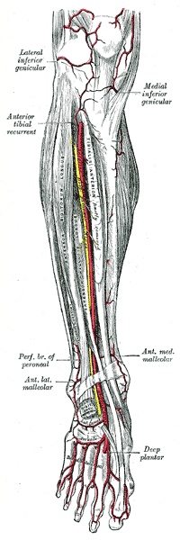
Dorsalis Pedis Artery
Original Editor - Priyanka Chugh
Top Contributors - Priyanka Chugh, Lucinda hampton, Nina Myburg, Kim Jackson, Joao Costa and Evan Thomas
Description[edit | edit source]
The dorsalis pedis artery (dorsal artery of foot), is a blood vessel of the lower limb that carries oxygenated blood to the dorsal surface of the foot. It arises at the anterior aspect of the ankle joint and is a continuation of the anterior tibial artery. It passes from the ankle joint along the tibial side of the dorsum of the foot to the proximal part of the intermetatarsal space. There it divides into two branches, the first dorsal metatarsal artery and the deep plantar artery. [1] Along its course, it is accompanied by a deep vein, the dorsalis pedis vein. The extensor hallucis longus and extensor digitorum brevis and longus tendons also can also be located close by.
Peculiarities in Size[edit | edit source]
The dorsal artery of the foot may be larger than usual, to compensate for a deficient plantar artery. It's terminal branches to the toes may be absent, the toes then being supplied by the medial plantar artery or its place may be taken altogether by a large perforating branch of the peroneal artery.
Palpation of the dorsalis pedis artery pulse[edit | edit source]
The dorsalis pedis artery pulse can be palpated lateral to the extensor hallucis longus tendon (or medially to the extensor digitorum longus tendon) on the dorsal surface of the foot, distal to the dorsal most prominence of the navicular bone which serves as a reliable landmark for palpation. It is often examined, by physicians, when assessing whether a given patient has peripheral vascular disease. It is absent, unilaterally or bilaterally, in 2–3% of young healthy individuals.[2]
Branches[edit | edit source]
The branches of the dorsalis pedis artery are:
- Lateral tarsal artery: (a. tarsea lateralis; tarsal artery) arises from the dorsalis pedis as it crosses the navicular bone. It passes in an arched direction lateralward, lying upon the tarsal bones, and covered by the Extensor digitorum brevis. It supplies this muscle and the articulations of the tarsus, and anastomoses with branches of the arcuate, anterior lateral malleolar and lateral plantar arteries, and with the perforating branch of the peroneal artery.
- Medial tarsal artery: (aa. tarseæ mediales) are two or three small branches on the medial border of the foot and join the medial malleolar network.
- Arcuate artery: (a. arcuata; metatarsal artery) arises anterior to the lateral tarsal artery. It passes lateralward, over the bases of the metatarsal bones, beneath the tendons of the Extensor digitorum brevis, it's direction being influenced by its point of origin, and it's anastomoses with the lateral tarsal and lateral plantar arteries. This vessel gives off the second, third, and fourth dorsal metatarsal arteries.
- First dorsal metatarsal artery: (a. dorsalis hallucis) runs forward on the first Interosseous dorsalis, and at the cleft between the first and second toes divides into two branches.
- Deep plantar artery: (ramus plantaris profundus; communicating artery) descends into the sole of the foot, between the two heads of the first Interosseous dorsalis, and unites with the termination of the lateral plantar artery, to complete the plantar arch. It sends a branch along the medial side of the great toe, and is continued forward along the first interosseous space as the first plantar metatarsal artery, which bifurcates for the supply of the adjacent sides of the great and second toes. [1]
Significance[edit | edit source]
Lack of a dorsalis pedis pulse may be due to peripheral vascular disease, hypovolemia, or cardiac dysfunction.
References[edit | edit source]
- ↑ 1.0 1.1 Gray H. Anatomy of the Human Body. Twentieth edition. Philadelphia: Lea & Febiger; 1918 Available from: https://www.bartleby.com/107/161.html [Accessed on 23 May 2019]
- ↑ Moore KL, Dalley AF. Clinically Oriented Anatomy. Fifth edition. Philadelphia: Lippincot Williams & Wilkins; 2006


