Diastasis Recti Abdominis
Original Editor - Marianne Ryan
Top Contributors - Nicole Hills, Sivapriya Ramakrishnan, Lucinda hampton, Admin, Vidya Acharya, Victoria Geropoulos, Regan Haley, Rachael Lowe, Laura Ritchie, Marianne Ryan, Oyemi Sillo, Kim Jackson, Michelle Walsh, WikiSysop, Tarina van der Stockt and Claire Knott - Your name will be added here if you are a lead editor on this page.
Definition/Description[edit | edit source]
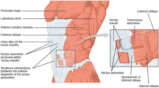
Diastasis recti abdominis is a separation of the rectus abdominal muscles at the linea alba.[2]
Diastasis recti abdominis can occur as result of prolonged transverse stresses on the linea alba in men,[3] postmenopausal women,[4] and in women during pregnancy. [5][6]
Diastasis recti can also occur in newborns and is defined as an inter-rectus distance that is greater than 3cm in this population.[7]
Diastasis Recti Abdominis and Pregnancy[edit | edit source]
During pregnancy, the linea alba must soften and expand to accommodate the growing fetus[8] increasing the width of the linea alba (the inter-rectus distance or IRD).
DRA is experienced by some (potentially most) women during their 3rd trimester of pregnancy.[8][9]
Clinically Relevant Anatomy[edit | edit source]
The two rectus abdominis muscle heads run parallel to each other and are separated by a connective tissue along the midline of the body called the linea alba.[10] The distance between the rectus abdominis muscles is referred to as the inter-rectus distance.
Clinical Presentation[edit | edit source]
An adult is considered to have a diastasis recti abdominis when they present with an 'abnormal' inter-rectus distance. On palpation, if a therapist can place two or more finger breaths (≈2cm) in the sulcus between the medial borders of the rectus abdominus muscles the patient may present with diastasis recti abdominis.[11] Palpation, in clinical practice, appears to be sufficient for detecting the presence or absence of diastasis recti,[12] however ultrasound imaging can produce a more precise measurement.[12]
Currently, the literature is not in agreement with what the diagnostic cut-off is for diastasis recti abdominis. Our best approach is to inform our clinical practices with the work done by Beer and colleagues[13] who suggest that, in nulliparous women (women who have not given birth), the normal width of the linea alba should be less than 1.5 cm at the xiphoid level, less than 2.2 cm at 3 cm above the umbilicus, and less than 1.6 cm at 2 cm below the umbilicus.[13]
Management / Interventions - Pregnancy and Postpartum[edit | edit source]
There is some evidence to support the idea that an increased inter-rectus distance is associated with the severity of self-reported abdominal pain.[15][16] There is no evidence to support the suggestion that there is an association between inter-rectus distance and lumbopelvic pain.[9][17]
Despite suggestions that diastasis recti abdominis may be associated with pelvic floor dysfunction, women who present with diastasis recti abdominis have not been shown to demonstrate pelvic floor muscle weakness or higher incident urinary incontinence or pelvic organ prolapse.[18]
A recent randomized control trial by Gluppe and colleagues[19] found that a weekly supervised postpartum exercise program and daily pelvic floor exercises did not reduce the prevalence of diastasis recti when compared to a control group.[19] The exercise program used in this study can be found here.
To date, there is currently not enough evidence in the literature to guide clinical practice on how and if we should 'close the gap' in women who present with diatasis recti abdominis after pregnancy.[19]
Patient Education[edit | edit source]
It is important to educate our patients on diastasis recti abdominis during and after pregnancy. The video below by a Canadian physiotherapist uses a great analogy to explain the concept of diastasis recti abdominis.
Common Postpartum Exercises[edit | edit source]
Kegel Exercises[edit | edit source]
Pelvic floor exercises can help strengthen deep abdominal muscles because all the core muscles contract automatically as a cohesive group. These exercises can be performed throughout pregnancy and may be started in the early postpartum period.
Breathing Exercises[edit | edit source]
Breathing exercises help to retrain the diaphragm to relearn how to descend after childbirth. During pregnancy, the diaphragm is pushed upwards by the growing uterus and loses its ability to descend during inhalation. Since the diaphragm forms the top of the core muscles, it is important to retrain it to function with a full excursion again. Here are 2 good exercises
- Inverted Breathing - Lie supine with the buttock on a pillow wedge, raised above chest level. Teach patient to take short, shallow breaths “through the pelvic diaphragm.” Have patient place one hand just above the pubic symphysis and teach them to feel for a slight up and down motion while they breathe in and out. Place the patient's other hand on top of their chest and tell them to try not to have their chest rise when breathing.
- Lateral Costal Breathing - In the seated position have patient place their hands on the lateral sides of the rib cage. Teach them to “breathe into their hands” and to feel for the lateral expansion of the rib cage as they inhale and the movement of the ribcage toward the midline during exhalation.
Progressive Core Exercises[edit | edit source]
There are several exercises that can be used to help restore core strength after pregnancy. For more information refer to Diane Lee’s work on the Diastasis Recti.
Precaution: Sit-Ups and Crunches[edit | edit source]
Sit-ups and similar exercises can put a strain on the pelvic floor and can increase intra-abdominal pressure which may not be helpful during pregnancy and when restoring the abdominal muscles and pelvic floor in the postpartum period.
Body Mechanics[edit | edit source]
It is important to teach the patient how to perform ADL’s without increasing abdominal pressure, such as rolling out of bed, instead of doing a “sit-up” to get up. Avoid “jack-knifing” behavior that increases intra-abdominal pressure. Teaching patients to roll to their left side and use their top arm to help push themselves up is a commonly prescribed technique. Other activities to watch out for is getting out of a bath, lifting and carrying older children and heavy objects during pregnancy and the early postpartum period. Teach them to “exhale as you lift.”
Postural Awareness[edit | edit source]
After pregnancy women tend to stand with and an exaggerated anterior pelvic tilt and with their pelvis pushed forward. In order to stand up against gravity, their bodies typically develop areas of rigidity in the upper lumbar and the lower thoracic area along with the buttock muscles. Diane Lee refers to this as “back clenching and buttock griping behavior.” Manual therapy and relaxation exercises may be indicated before initiating strengthening exercises. (http://dianelee.ca/articles/UnderstandYourBack&PGPopt.pdf)
Abdominal Supports[edit | edit source]
Abdominal Binders: Binders may be helpful for some women in the postpartum period, but the wrong or overuse of them can cause more problems. It is best not to use an abdominal binder unless necessary, for example during pregnancy in the third trimester and 6 weeks after delivery if there is a separation of 2 fingers or more.
Belly Band: Wearing a belly band during pregnancy (one made specifically for pregnant women) may help increase proprioception and muscle awareness. A belly band or tight tube top can be used after childbirth for the same reasons.
References[edit | edit source]
- ↑ Wikimedia Commons.https://commons.wikimedia.org/wiki/File:1112_Muscles_of_the_Abdomen_Anterolateral.png (accessed 22 June 2018).
- ↑ Gilleard WL, Brown JM. Structure and function of the abdominal muscles in primigravid subjects during pregnancy and the immediate postbirth period. Physical therapy. 1996 Jul 1;76(7):750-62.
- ↑ Lockwood T. Rectus muscle diastasis in males: primary indication for endoscopically assisted abdominoplasty. Cosmetic. 1998;May:1685-1691.
- ↑ Spitznagle TM, Leong FC, Van Dillen LR. Prevalence of diastasis recti abdominis in a urogynecological patient population. International Urogynecology Journal. 2007 Mar 1;18(3):321-8.
- ↑ Akram J, Matzen SH. Rectus abdominis diastasis. Journal of plastic surgery and hand surgery. 2014 Jun 1;48(3):163-9.
- ↑ Brauman D. Diastasis recti: Clinical anatomy. Plastic and reconstructive surgery. 2008 Nov 1;122(5):1564-9.
- ↑ Marden PM, Smith DW, McDonald MJ. Congenital anomalies in the newborninfant, including minor variations: A study of 4,412 babies by surface examination for anomalies and buccal smear for sex chromatin. The Journal of pediatrics. 1964 Mar 1;64(3):357-71.
- ↑ 8.0 8.1 Boissonnault JS, Blaschak MJ. Incidence of diastasis recti abdominis during the childbearing year. Physical therapy. 1988 Jul 1;68(7):1082-6.
- ↑ 9.0 9.1 Mota PG, Pascoal AG, Carita AI, Bø K. Prevalence and risk factors of diastasis recti abdominis from late pregnancy to 6 months postpartum, and relationship with lumbo-pelvic pain. Manual therapy. 2015 Feb 1;20(1):200-5.
- ↑ Peterson-Kendall F, Kendall-McCreary E, Geise-Provance P, McIntyre-Rodgers, Romani W. Muscles: Testing and Function, with Posture and Pain. 5th ed. Baltimore: Wolters Kluwer; 2005.
- ↑ Noble E. Essential Exercises for the Childbearing Year. 2nd editio. Boston, MA: Houghton Miffilin; 1982.
- ↑ 12.0 12.1 Van de Water AT, Benjamin DR. Measurement methods to assess diastasis of the rectus abdominis muscle (DRAM): a systematic review of their measurement properties and meta-analytic reliability generalisation. Manual therapy. 2016 Feb 1;21:41-53.
- ↑ 13.0 13.1 Beer GM, Schuster A, Seifert B, Manestar M, Mihic‐Probst D, Weber SA. The normal width of the linea alba in nulliparous women. Clinical anatomy. 2009 Sep 1;22(6):706-11.
- ↑ Learn with Diane Lee. Linea alba screen DRA with Diane Lee. Available from: https://www.youtube.com/watch?v=06o8Z54l-40 [last accessed 22/06/2018]
- ↑ Parker MA, Millar LA, Dugan SA. Diastasis Rectus Abdominis and Lumbo‐Pelvic Pain and Dysfunction‐Are They Related?. Journal of Women’s Health Physical Therapy. 2009 Jul 1;33(2):15-22.
- ↑ Keshwani N, Mathur S, McLean L. Relationship Between Inter-rectus Distance and Symptom Severity in Women With Diastasis Recti in the Early Postpartum Period. Physical therapy. 2017 Dec 4.
- ↑ Sperstad JB, Tennfjord MK, Hilde G, Ellström-Engh M, Bø K. Diastasis recti abdominis during pregnancy and 12 months after childbirth: prevalence, risk factors and report of lumbopelvic pain. Br J Sports Med. 2016 Jun 20:bjsports-2016.
- ↑ Bø K, Hilde G, Tennfjord MK, Sperstad JB, Engh ME. Pelvic floor muscle function, pelvic floor dysfunction and diastasis recti abdominis: Prospective cohort study. Neurourology and urodynamics. 2017 Mar 1;36(3):716-21.
- ↑ 19.0 19.1 19.2 Gluppe SL, Hilde G, Tennfjord MK, Engh ME, Bø K. Effect of a Postpartum Training Program on Prevalence of Diastasis Recti Abdominis in Postpartum Primiparous Women: A Randomized Controlled Trial. Physical therapy. 2018 Jan 17.
- ↑ Phit Physiotherapy. DRA: Being (banana) split up the middle, a fresh (produce) perspective. Available from: https://www.youtube.com/watch?v=rVxAUOkb3M4[last accessed 22/06/2018]
- ↑ Diane Lee's Integrated Systems Model for Physiotherapy in Womens' Health. Available from:https://www.youtube.com/watch?v=5oslM6Pe9AU&t=1844s [last accessed 22/6/2018]
- ↑ Diane Lee. Conference Presentations Diane Lee and Associates in Physiotherapy. Available from: https://www.youtube.com/watch?v=mHY6CSSosNE&t=10s[last accessed 22/6/2018]


