Current Management of Lisfranc Injuries
Top Contributors - Ewa Jaraczewska and Jess Bell
Definition and Epidemiology[edit | edit source]
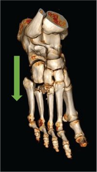
The Lisfranc joint is where the tarsal bones connect to the metatarsal bones - i.e. the tarsometatarsal (TMT) joint. A 'Lisfranc injury' was first described in 1815 by the French surgeon and gynaecologist Jacques Lisfranc de St. Martin (1787- 1847). It is a condition where one or more of the metatarsal bones are displaced.[1]
According to Ponkilainen et al.,[2] the epidemiology of mid-foot injuries, including Lisfranc injuries, is not well known. A five-year study found that out of all mid-foot traumas, Lisfranc injury occurred in 18.2% of cases. A survey by Desmond and Chou[3] confirmed this type of injury in less than 2% of all fractures. However, statistics suggest that diagnosis is not always adequate. Late or missed diagnosis occurs in 20% of cases.[4] It has been found that:
- Lisfranc injuries are more common in men, and the reported ratio is 4.25:1.[5]
- The most common cause of traumatic injury is low-energy trauma[6] (e.g. falling over a plantar flexed foot).[7]
- The most frequent high-energy trauma is a car accident, followed by a motorcycle accident.[5]
- The most common fractures include the second and fifth metatarsal bones,[8] followed by the cuboid bone.[5]
- Multiple metatarsal fractures are not uncommon. In these cases, fractures of the third metatarsal are associated with a fracture of either the second or fourth metatarsal.[8]
- Lisfranc injuries occur more frequently in athletes.[9]
Risk Factors[edit | edit source]
The following can increase the risk of an unstable injury:[6]
- Intraarticular fractures in the two lateral tarsometatarsal joints
- Female gender
- A shorter height of the second tarsometatarsal joint
Clinically Relevant Anatomy[edit | edit source]
Lisfranc injuries involve trauma to the tarsometatarsal (TMT) joint. The descriptions of this injury vary significantly, as does the documentation of the anatomy of the Lisfranc joint.[10]
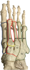
The Lisfranc joint complex includes the first to fifth metatarsal bones, three cuneiform bones, the cuboid, the interconnecting ligaments, capsules, and reinforcing tendons. The primary role of this joint is to provide mid-foot stability.[11] The metatarsal bases create an arch structure, with the second metatarsal bone as its midpoint (i.e. keystone).[11] It articulates with the second cuneiform bone and sits between the first and third cuneiforms. The following tarsometatarsal joint ligaments add to the strength of the joint:[11]
- Dorsal ligaments (longitudinal direction)
- Interosseous ligaments (transverse direction)
- Plantar ligaments (oblique direction)
There are three tarsometatarsal synovial joint systems that relate to the three columns of the foot:[11]
- Medial column (first cuneiform bone to the first metatarsal bone)
- Middle column (second cuneiform bone to the second metatarsal and third cuneiform to the third metatarsal)
- Lateral column (cuboid bone to the fourth and fifth metatarsal bones)
You can read about the foot bones and ligaments here.
Mechanism of Injury / Pathological Process[edit | edit source]
Lisfranc injuries can be caused by:
- Direct, high level of energy: crush injury to the joint region, industrial accident (heavy weight lands on foot when dropped from a height), fall from a height, motor vehicle accident.[9]
- Indirect, low level of energy: sports injury that "usually involves a longitudinal force while the foot is plantar flexed with a medial or lateral rotational force",[9] stepping off a curb with the foot forcefully plantar flexed,[12] ground-level fall.
Classification of Lisfranc injuries includes:[13]
- Traditional dislocation (first and second tarsometatarsal ligament tear)
- Medial column dislocation (second tarsometatarsal and medial-middle cuneiform ligament tear)
- Proximal extension dislocation (first, second, and medial-middle cuneiform ligament tear)
Low energy injuries include partial injuries (sprains). In this type of injury, the plantar tarsometatarsal ligaments remain intact, and the injury is stable.[14]
Diagnosis[edit | edit source]
Assessment[edit | edit source]
Each assessment should include the following components:[14]
- An interview/medical history
- Exact injury mechanism, including the position of the foot, the direction of force, and the extent of energy involved
- Location of the pain and potential structures involved
- Palpation of the midfoot
- Soft tissue inspection
- Checking for plantar ecchymosis (bruising) at the midfoot
- Provocative manoeuvres to evaluate for instability: squeezing of the midfoot, pronation, supination, abduction, adduction, single limb weight bearing
- Passive range of motion in the sagittal and coronal planes of all three columns of the foot
Clinical Presentation[edit | edit source]
- Diffuse pain in the mid-foot, usually lasting for a few days. Pain refers to the third toe when pressure is applied at the mid-foot [7]
- Tenderness to palpation of the mid-foot[14]
- Reproduction of pain with passive motion of the forefoot[14]
- Inability to weight bear[14] / difficulties weight bearing
- Development of compensatory strategies for pain and mobility restrictions, including wearing stiff leather shoes, or boots[7]
- Pitting oedema[7]
- Flattening of the transverse arch[7]
- Plantar ecchymosis at the midfoot due to ligament rupture[14]
Diagnostic Procedures[edit | edit source]
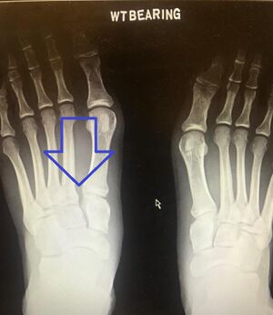
- Radiography to evaluate diastasis at the TMT joints includes:[11]
- Standard, three-view films of the foot—anteroposterior, 30-degree internal oblique, and lateral views.
- The patient should be standing for all films, with weight-bearing.
- An x-ray should be completed of both feet for comparison.
- Radiographic images completed in non-weight-bearing positions tend to miss Lisfranc injuries 20-50% of the time.
According to Yan et al.,[11] the diagnostic accuracy of radiographs is low. If clinical symptoms continue, but the X-ray is negative, the patient should be referred for computed tomography (CT) or magnetic resonance imaging (MRI).[11]
- CT to detect non-displaced fractures and minimal osseous subluxation.[15]
- MRI to evaluate ligament abnormalities.[15]
Recommendations for Intervention[11][edit | edit source]
- Conservative management:
- Lisfranc injuries without evidence of instability (<2 mm diastasis)
- Extra-articular fractures with stability confirmed by weight-bearing[14]
- Operative management:
- All types of injury not listed above as suitable for surgical management
- There is a lack of one optimal surgical treatment
- There is also a lack of concrete evidence supporting any particular treatment modality
- All types of injury not listed above as suitable for surgical management
Management / Intervention[edit | edit source]
Conservative Management[7][edit | edit source]
- Pain management
- Medial arch support. The type of support depends on the degree of injury and can include: wearing a boot, supportive shoes or a customised orthotic
- Immobilisation: immobilise in a cast or boot for 6-10 weeks to offload the foot
- Limit weight-bearing activities: do not push for full weight bearing
- Arch support: consider arch support in the boot and the cast
- Strengthening programme for:
- Intrinsics of the foot to improve dynamic foot support
- Toe flexors to help with toe push off
- Tibialis posterior
- Isometric strengthening for peroneus longus
- Taping the metatarsal arch to improve stability
- Cardiovascular fitness
- Core strengthening
Surgical Management[edit | edit source]
When the injury is unstable, surgical management for the ligaments, bone or a combination of both is indicated:[14]
- Reduction and immobilisation of any dislocation-produciong tension of the skin and soft tissue.[14]
- Temporary external fixation for high-energy injuries is indicated when the splint is insufficient to maintain alignment.[14]
- Tissue swelling must be resolved before definitive surgery is scheduled (10-14 days after injury).[14] The general rule for the timing of the surgery is "the sooner, the better as long as the swelling is manageable."[11]
- Open reduction and internal fixation (ORIF) with transarticular screw fixation.[14]
- ORIF with primary arthrodesis when a pure ligamentous pattern is present.[14]
Good anatomical reduction in the surgery significantly improves the outcome.[7] Helene Simpson
Resources[edit | edit source]
- Paek S, Mo M, Hogue G. Treatment of paediatric Lisfranc injuries: A systematic review and introduction of a novel treatment algorithm. Journal of Children's Orthopaedics. 2022 May 10:18632521221092957.
- van den Boom NA, Stollenwerck GA, Lodewijks L, Bransen J, Evers SM, Poeze M. Lisfranc injuries: fix or fuse? a systematic review and meta-analysis of current literature presenting outcomes after surgical treatment for Lisfranc injuries. Bone & Joint Open. 2021 Oct 8;2(10):842-9.
- Lakey E, Hunt KJ. Patient-Reported Outcomes in Foot and Ankle Orthopedics. Foot Ankle Orthop. 2019 Jul 19;4(3):2473011419852930.
References[edit | edit source]
- ↑ Moracia-Ochagavía I, Rodríguez-Merchán EC. Lisfranc fracture-dislocations: current management. EFORT Open Rev. 2019 Jul 2;4(7):430-444.
- ↑ Ponkilainen VT, Laine HJ, Mäenpää HM, Mattila VM, Haapasalo HH. Incidence and characteristics of midfoot injuries. Foot & Ankle International. 2019 Jan;40(1):105-12.
- ↑ Desmond EA, Chou LB. Current concepts review: Lisfranc injuries. Foot Ankle Int. 2006 Aug;27(8):653-60.
- ↑ Myerson MS, Cerrato R. Current management of athletes' tarsometatarsal injuries. Instr Course Lect. 2009;58:583-94.
- ↑ 5.0 5.1 5.2 Sobrado MF, Saito GH, Sakaki MH, Pontin PA, Santos AL, Fernandes TD. Epidemiological study on Lisfranc injuries. Acta Ortopédica Brasileira. 2017 Jan;25:44-7.
- ↑ 6.0 6.1 Stødle AH, Hvaal KH, Enger M, Brøgger H, Madsen JE, Husebye EE. Lisfranc injuries: incidence, mechanisms of injury and predictors of instability. Foot and Ankle Surgery. 2020 Jul 1;26(5):535-40.
- ↑ 7.0 7.1 7.2 7.3 7.4 7.5 7.6 Simpson H. Lisfranc Injuries Course. Plus, 2022.
- ↑ 8.0 8.1 Petrisor BA, Ekrol I, Court-Brown C. The epidemiology of metatarsal fractures. Foot & ankle international. 2006 Mar;27(3):172-4.
- ↑ 9.0 9.1 9.2 Buchanan BK, Donnally III CJ. Lisfranc dislocation. InStatPearls [Internet] 2021 Jul 25. StatPearls Publishing.Last Update: February 2, 2022.
- ↑ DeLuca MK, Boucher LC. Morphology of the Lisfranc Joint Complex. The Journal of Foot and Ankle Surgery. 2022 Jul 25.
- ↑ 11.0 11.1 11.2 11.3 11.4 11.5 11.6 11.7 11.8 Yan A, Chen SR, Ma X, Shi Z, Hogan M. Updates on Lisfranc Complex Injuries. Foot Ankle Orthop. 2021 Jan 25;6(1):2473011420982275.
- ↑ Kalia V, Fishman EK, Carrino JA, Fayad LM. Epidemiology, imaging, and treatment of Lisfranc fracture-dislocations revisited. Skeletal Radiol. 2012 Feb;41(2):129-36.
- ↑ Porter DA, Barnes AF, Rund A, Walrod MT. Injury pattern in ligamentous Lisfranc injuries in competitive athletes. Foot & Ankle International. 2019 Feb;40(2):185-94.
- ↑ 14.00 14.01 14.02 14.03 14.04 14.05 14.06 14.07 14.08 14.09 14.10 14.11 14.12 Clare MP. Lisfranc injuries. Curr Rev Musculoskelet Med. 2017 Mar;10(1):81-85.
- ↑ 15.0 15.1 Sripanich Y, Weinberg MW, Krähenbühl N, Rungprai C, Mills MK, Saltzman CL, Barg A. Imaging in Lisfranc injury: a systematic literature review. Skeletal Radiology. 2020 Jan;49(1):31-53.
- ↑ Sandringham Sports Physio. Foot Arch Supportive Taping.2019. Available from: https://www.youtube.com/watch?v=7QM41olCNS4 [last accessed 29/09/2022]


