Colles Fracture: Difference between revisions
No edit summary |
mNo edit summary |
||
| Line 27: | Line 27: | ||
== Differential diagnosis == | == Differential diagnosis == | ||
*[[Image: | *[[Image:Colles fracture.JPG|thumb|right]]Radiographic Imaging - dorsally angulated fracture of distal radial metaphysis | ||
*CT Scan | *CT Scan | ||
Revision as of 07:00, 10 May 2017
Original Editor - Stacy S Stone
Top Contributors - Emma Guettard, Admin, Adam Vallely Farrell, Kim Jackson, Stacy S Stone, Laura Ritchie, Rachael Lowe, Anne-Laure Vanherwegen, Nikhil Benhur Abburi, Lauren Lopez, Lucinda hampton, Scott Buxton, Priyanka Chugh, Benjamin Desmedt, WikiSysop, Claire Knott, Anas Mohamed and Evan Thomas - Emma Guettard as part of the Vrije Universiteit Brussel Evidence-based Practice Project
Definition/Description[edit | edit source]
A colles fracture is a fracture of the distal radius. It was first described in 1814, by Abraham Colles, an Irish surgeon. The fracture originates from a fall on the outstretched hand and is usually associated with dorsal and radial displacement of the distal fragment, and disturbance of the radial-ulnar articulation. Possibly the ulnar styliod may be fractured. Communication of the distal fragment and fractures into the joint surface are present in some of these fractures. The colles fracture is one of the most common and challenging of the outpatient fractures[1]. Colles' fracture is defined as a linear transverse fracture of the distal radius approximately 20-35 mm proximal to the articular surface with dorsal angulation of the distal fragment[2].
Clinical relevant anatomy
[edit | edit source]
Low energy extra-articular fracture of the distal radius. Can be associated with ulnar styloid fracture, TFCC tear, scapholunate dissociation.[8]
Epidemiology/Etiology [edit | edit source]
Females are predilected more than males for this type of injury and oftentimes there is a precedent history of osteoporosis.' In the United States and in Northern Europe, colles fractures are the most common fractures in women up to the age of 75 years[3]. It is known that these fractures appear mostly by young adults and the elderly[4]. Stable Colles' fractures present with minimal comminution. Unstable fractures are distinctly comminuted often with corresponding avulsions of the radial or ulnar styloid, that have the potential to cause compression neuropathies, especially of the median nerve. Other complications that have been reported are degenerative joint disease and reflex sympathetic dystrophy[2].
Characteristics/clinical presentation
[edit | edit source]
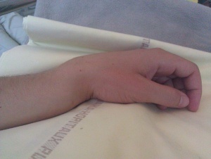
- "Dinner Fork" Deformity[5]
- History of fall on an outstretched hand
- Dorsal wrist pain
- Swelling of the wrist
- Increased angulation of the distal radius
- Inability to grasp object[6]
Differential diagnosis [edit | edit source]
- Radiographic Imaging - dorsally angulated fracture of distal radial metaphysis
- CT Scan
Classifications of Distal Radial (Colles') Fracture
- Universal Classification of Dorsally Displaced Distal Radial Fractures Type I - undisplaced
- Universal Classification of Dorsally Displaced Distal Radial Fractures Type II - displaced
- Melone Type I - undisplaced and minimally comminuted
- Frykman Type I - distal radial fracture without distal ulnar fracture
- Frykman Type II - distal radial fracture with distal ulnar fracture[7]
Outcome Measures[edit | edit source]
Examination [edit | edit source]
Medical management [edit | edit source]
| [12] |
There are a number of options for stabilization and medical treatment of this fracture. These comprise conservative management with cast immobilization or surgical options: external fixation, internal fixation, percutaneous pinning, and bone substitutes. The fracture pattern, degree of displacement, the stability of the fracture, and the age and physical demands of the patient provide the best treatment option.
Conservative Treatment
- Immobilization in cast/splint - typically positioned in slight flexion, pronation
- Percutaneous Pinning
Surgical Intervention
- ORIF[7]
Physical therapy [edit | edit source]
Rehabilitation protocol for Colles’ fracture[edit | edit source]
Passive:[edit | edit source]
A case report used a rehabilitation protocol to improve range of motion and grip strength in an undisplaced, stable Colles' fracture. The patient got a treatment with passive interventions to improve circulation and prevent immobilization adhesion formation. These treatments included application of an ice pack to reduce edema followed by application of a wax bath on the affected wrist. Gentle range of motion mobilizations were then introduced that could only be performed in flexion and extension to the patient's pain tolerance. Three sets of 5 flexion/extension repetitions were performed on the affected wrist. The joint was also mobilized in circumduction, ulnar flexion and radial flexion to the patient's level of tolerance.
Early mobilisation resulted in rapid recovery of both movement and strength without causing more discomfort or adversely influencing the progression of the deformity. In patients over 55, minimally displaced fractures can safely be treated in a crepe bandage, and displaced fractures which have been reduced can be treated in a modified cast. Early mobilisation would ensure rapid recovery of wrist and hand function while avoiding the complications of a conventional plaster cast[13].
This study [13] proved that in the groups with displaced and undisplaced fractures, the recovery of forearm rotation and finger movement paralleled the recovery of wrist movement: for both types of fracture, early mobilisation led to an earlier return of strength. Although this recovery did not parallel the improvement in wrist movement.
In both categories early mobilization led to more rapid resolution of wrist swelling in the first five weeks. At nine weeks and at 13 weeks the wrist girths were similar.
Patients encouraged to mobilise the injured wrist from the outset recovered wrist movement more quickly than those who were imobilised in a conventional plaster cast.[13]
Supervised Active rehabilitation program:[edit | edit source]
- ISOMETRIC EXERCISE
- wrist flexors and extensors
- ACRIVE RANGE OF MOTION EXERCISE
- assisted strech to forearm flexors ans extensor musculature ans radial/ulnar deviation
- weight bearing wrist extension exercise(hand on the table with the patient leaning forward on them) topatient tolerance
- active strech to shoulder girdle and rotator cuff musculature
- active strech to elbow flexor and extensor musculature
- INTRINSIC HAND MUSCLE EXERCISE
- thumb/digit opposition
- repetitive squeezing of theraputty
- repetitive towel wringing exercise
- STRENGTHENING ROUTINE
- biceps curl with 1,5-2 pound weights bilaterally
- shoulder abduction, flexion and extension reps with 2 pound weights bilaterally
- repetitive squeezing of rubber ball in affected wrist
- flexion ans extension of wrist using 1,5 pound weights increasing as tolerated
- FUNCTIONAL ACTIVITIES
- patient is encouraged to resume pre-accident activities that involve the affected extremity (eg. writing, typing, cooking, etc.)
In addition the patient in this case study[2] was encouraged to resume functional activities that involve the wrist and hand such as writing, cooking and sewing.
In the study of Gupta A. fractures immobilised with the wrist in dorsiflexion had the best results.
Comparison of various joint movements with those on the uninjured side showed that fractures who were immobilised in palmar flexion had more joint stiffness, particularly of the metacarpophalangeal and interphalangeal joints. Even in type 3 fractures (displaced,extra-articular with communition), where the position of immobilization of the wrist did not significantly effect the anatomical result, immobilization in dorsiflexion provided the best recovery of function[14].
In the study[15] it is proved that the addition of occupational therapy to instruction therapy reaches no statistical significance for following variables. Parameters examined were the dorsal angulation, the radial angulation and the axial radial length.
Functional scores were measured such as the modified Gartland and Werley score, but there were no statistical differences.
The instruction therapy consists of the following activities: a warm-up for 10 min in warm soap-water, raising and lowering the shoulders, rotating the shoulders, bending and stretching the fingers, spreading and joining the fingers, reaching the fingertips with the thumb, reaching the finger base with the thumb, turning the back and palm of the hand with the elbow fixed at the side and finally bending the wrist with the hand over the side of a table. The occupational therapy includes active joint exercises for wrist, elbow and shoulder, edema prevention, coordination exercise, coarse and fine motor-function exercise, strengthening exercise, sensation exercise and ADL training[15].
Key Research [edit | edit source]
- Arora R, Gabl M, Gschwentner M, Deml C, Krappinger D, Lutz M. A comparative study of clinical and radiologic outcomes of unstable colles type distal radius fractures in patients older than 70 years: nonoperative treatment versus volar locking plating. J Orthop Trauma. 2009;23(4):237-42. (level of evidence 2b)
- Wright TW, Horodyski M, Smith DW. Functional outcome of unstable distal radius fractures: ORIF with a volar fixed-angle tine plate versus external fixation. J Hand Surg Am. 2005;30(2):289-99.(level of evidence 1b)
- Handoll HH, Huntley JS, Madhok R. External fixation versus conservative treatment for distal radial fractures in adults. Cochrane Database Syst Rev. 2007 Jul 18;(3):CD006194.(level of evidence 1a)
- Gehrmann SV, Windolf J, Kaufmann RA. Distal radius fracture management in elderly patients: a literature review. J Hand Surg Am. 2008;33(3):421-9.(level of evidence 2a)
Recent Related Research (from Pubmed)[edit | edit source]
Failed to load RSS feed from http://www.ncbi.nlm.nih.gov/entrez/eutils/erss.cgi?rss_guid=1naYwjERihpqD2ZPYSOhhDP07m6EgLs4B0iIPvXCGObwdflQIf|charset=UTF-8|short|max=10: Error parsing XML for RSS
References[edit | edit source]
- ↑ T. M. Molder, E. Vernon Stabler, M.D., and William H. Cassebaum,M.D.; Colles fracture: evaluation and selection of the therapy; the journal of trauma and acute case surgery. 1965; volume 5 issue 4.(Level of Evidence 1B)
- ↑ 2.0 2.1 2.2 Stephen Balsky, Rehabilitation protocol for undisplaced Colles' fractures following cast removal, the journal of the Canadian chiropractic association.(Level of evidence 4)
- ↑ Owen RA, Melton LJ, 3rd, Johnson KA, Ilstrup DM, Riggs BL., Incidence of Colles’ fracture in a North American community. Am J Public Health. 1982;72(6):605–607.
- ↑ Cummings SR, Kelsey JL, Nevitt MC, O’Dowd KJ., Epidemiology of osteoporosis and osteoporotic fractures. Epidemiol Rev. 1985;7:178–208.
- ↑ Hoynak BC, Hopson L. EMedicine. Wrist Fractures.http://emedicine.medscape.com/article/828746-overview (Acessed 2 July 2009).
- ↑ Joseph TN. Medline Plus. Colles' Wrist Fracture.http://www.nlm.nih.gov/medlineplus/ency/article/000002.htm (Accessed 2 July 2009).
- ↑ 7.0 7.1 Wheeless CR. Wheeless' Textbook of Orthopaedics. Colles Fracture. http://www.wheelessonline.com/ortho/colles_frx(Accessed 2 July 2009)
- ↑ MacDermid JC, Roth JH, Richards RS. Pain and disability reported in the year following a distal radius fracture: a cohort study. BMC Musculoskeletal Disord. 2003;4:24.
- ↑ Arora R, Gabl M, Gschwentner M, Deml C, Krappinger D, Lutz M. A comparative study of clinical and radiologic outcomes of unstable colles type distal radius fractures in patients older than 70 years: nonoperative treatment versus volar locking plating. J Orthop Trauma. 2009;23(4):237-242.
- ↑ Wright TW, Horodyski M, Smith DW. Functional outcome of unstable distal radius fractures: ORIF with a volar fixed-angle tine plate versus external fixation. J Hand Surg Am. 2005;30(2):289-299.
- ↑ Tremayne A, Taylor N, McBurney H, Baskus K. Correlation of impairment and activity limitation after wrist fracture. Physiother Res Int. 2002;7(2):90-99.
- ↑ besthandsurgeon. Distal Radius Fracture ORIF. Available from: http://www.youtube.com/watch?v=Ye839BYoMaY[last accessed 22/03/13]
- ↑ 13.0 13.1 13.2 Dias JJ, Wray CC, Jones JM, Gregg PJ.,The value of early mobilisation in the treatment of Colles' fractures. 1987;69(3); 463-7. (level of evidence 1a)
- ↑ Gupta A., The treatment of Colles' fracture. Immobilisation with the wrist dorsiflexed. 1991;73(2):312-5.(level of evidence 1b)
- ↑ 15.0 15.1 O.M. Christensen (2000) Occupational therapy and Colles’ fractures, International Orthopaedics (SICOT). 2001;25:43–45 (level of evidence 1b)


