Cerebellum
Introduction[edit | edit source]

The cerebellum is a vital component in the human brain as it plays a role in motor movement regulation and balance control. The cerebellum [1] (see image on R, horizontal fissure marked red)
- Coordinates gait
- Maintains posture,
- Controls muscle tone and voluntary muscle activity
- Is unable to initiate muscle contraction.
Damage to this area in humans results in a loss in the ability to control fine movements, maintain posture, and motor learning
The cerebellum is neuron-rich, containing 80% of the brain’s neurons organized in a dense cellular layer, and it's surface area when unfolded is nearly 75% of the surface area of the cerebrum.[1]
Anatomical Position[edit | edit source]
The cerebellum is located at the back of the brain, immediately inferior to the occipital and temporal lobes, and within the posterior cranial fossa. It is separated from these lobes by the tentorium cerebelli, a tough layer of dura mater.
It lies at the same level of and posterior to the pons, from which it is separated by the fourth ventricle.
Structure[edit | edit source]
The cerebellum consists of two hemispheres which are connected by the vermis, a narrow midline area. The cerebellum consists of grey matter and white matter:[2]
- Grey matter – located on the surface of the cerebellum. It is tightly folded, forming the cerebellar cortex. The gray matter of the cortex divides into three layers: an external - the molecular layer; a middle - the Purkinje cell layer; and an internal - the granular layer. The molecular layer contains two types of neurons: the outer stellate cell and the inner basket cell.[1]
- White matter – located underneath the cerebellar cortex. Embedded in the white matter are the four cerebellar nuclei (the dentate, emboliform, globose, and fastigi nuclei).
There are three ways that the cerebellum can be subdivided – anatomical lobes, zones and functional division[2]
- Anatomical Lobes
There are three anatomical lobes that can be distinguished in the cerebellum. These lobes are divided by two fissures – the primary fissure and posterolateral fissure;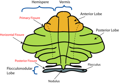
- The anterior lobe,
- The posterior lobe
- The flocculonodular lobe. It is the oldest part of the brain in evolutionary terms (archicerebellum) and participates mainly in balance and spatial orientation. Its primary connections are with the vestibular nuclei, although it also receives visual and other sensory input.[3]
- Zones
There are three cerebellar zones. In the midline of the cerebellum is the vermis. Either side of the vermis is the intermediate zone. Lateral to the intermediate zone are the lateral hemispheres. There is no difference in gross structure between the lateral hemispheres and intermediate zones - Functional Divisions
The cerebellum can also be divided by function. There are three functional areas of the cerebellum – the cerebrocerebellum, the spinocerebellum and the vestibulocerebellum.- Cerebrocerebellum – the largest division, formed by the lateral hemispheres. It is involved in planning movements and motor learning. It receives inputs from the cerebral cortex and pontine nuclei, and sends outputs to the thalamus and red nucleus. This area also regulates coordination of muscle activation and is important in visually guided movements.
- Spinocerebellum – comprised of the vermis and intermediate zone of the cerebellar hemispheres. It is involved in regulating body movements by allowing for error correction. It also receives proprioceptive information.
- Vestibulocerebellum – the functional equivalent to the flocculonodular lobe. It is involved in controlling balance and ocular reflexes, mainly fixation on a target. It receives inputs from the vestibular system, and sends outputs back to the vestibular nuclei.

Nerves[edit | edit source]
The cerebellum attaches to the brainstem by three groups of nerve fibers called the superior, middle and inferior cerebellar peduncles, through which efferent and afferent fibers pass to connect with the rest of the nervous system.
Function[edit | edit source]
Function by regions
- The cortex of the vermis coordinates the movements of the trunk, including the neck, shoulders, thorax, abdomen, and hips.
- Control of the distal extremity muscles is by the intermediate zone of cerebellar hemispheres, located adjacent to the vermis.
- The remaining lateral area of each cerebellar hemisphere provides the planning of sequential movements of the entire body along with involvement in the conscious assessment of movement errors
Main Functions overall[3]

- The cerebellum is essential for making fine adjustments to motor actions. Cerebellar dysfunction primarily results in problems with motor control.
- Four principles are important to cerebellar processing: feedforward processing, divergence and convergence, modularity, and plasticity.
- Signal processing in the cerebellum is almost entirely feedforward. Signals move through the system from input to output with very little internal transmission.
- The cerebellum both receives input and transmits output via a limited number of cells.
- The cerebellar system is divided into thousands of independent modules with similar structure.
Blood Supply[edit | edit source]

The cerebellum receives its blood supply from three paired arteries (originate from the vertebrobasilar anterior system)[2]
- Superior cerebellar artery (SCA)
- Anterior inferior cerebellar artery (AICA)
- Posterior inferior cerebellar artery (PICA)
The SCA and AICA are branches of the basilar artery, which wraps around the anterior aspect of the pons before reaching the cerebellum. The PICA is a branch of the vertebral artery.
Venous drainage of the cerebellum is by the superior and inferior cerebellar veins. They drain into the superior petrosal, transverse and straight dural venous sinuses.
Clinical Significance[edit | edit source]
The cerebellum receives afferent information about voluntary muscle movements from the cerebral cortex and from the muscles, tendons, and joints. It also receives information concerning balance from the vestibular nuclei. Each cerebellar hemisphere controls the same side of the body, thus if damaged the symptoms will occur ipsilaterally.[1]
Dysfunction of the cerebellum can produce a wide range of symptoms and signs.
The most common cause of cerebellar dysfunction is alcohol poisoning, but also trauma, multiple sclerosis, tumors, thrombosis of the cerebellar arteries and stroke.[1]
The clinical picture depends on the functional area of the cerebellum that is affected[3].
- Damage to the flocculonodular lobe (vestibulocerebellum): loss of equilibrium causing an altered walking gait
- Lateral zone damage: problems with skilled voluntary and planned movements leading to errors in intended movements (eg., dysdiadochokinesia, the inability to perform rapid alternating movements).
- Damage to the midline portion: disruption of whole-body movements
- Damage to the upper part of the cerebellum: gait impairments and other problems with leg coordination (ie, ataxia).
A wide variety of manifestations are possible (remembered by acronym ‘DANISH‘)[2]
- Dysdiadochokinesia - the lack of ability to perform rapidly alternating movements. Ask the patient to quickly supinate and pronate both forearms simultaneously. Movements will be slow and incomplete on the side of the cerebellar lesion.[1]
- Ataxia - voluntary movement disturbance involves tremor with fine movements eg writing or buttoning the clothes. Finger to nose test is performed to examine the coordination of the muscle movements, testing tip of the nose with the index finger test the movements are not properly coordinated, and tremor is observed at the end of the movement, called intention tremor. A similar test can be performed on the lower limbs ask patient to place the heel of one foot against the shin of the opposite leg.[1]
- Nystagmus (coarse) - Ataxia of ocular muscles, a rhythmical oscillation of the eyes. To provoke nystagmus, the patient should rotate eyes horizontally
- Intention tremor
- Dysarthria/Scanning speech - ataxia of the larynx muscles, speech is slurred and syllables are separated from one another.
- Hypotonia - the muscles lose resistance to palpation due to diminished influence of the cerebellum on gamma motor neurons. The patient walks with a broad-based gait and leans toward the affected side.[1]
Occlusion of PICA cause Wallenberg syndrome
The short video below gives a good picture of the manifestations of cerebellum damage
The cerebellum appears to play a role in many types of behaviours. Cerebellar damage not only affects movement coordination, but also disrupts some perceptual abilities such as visual motion discrimination. The cerebellum acts to make predictions for different cerebral areas of the brain to optimize their abilities-- helping to predict optimal motor commands for movement control and upcoming sensory events for sensory perception possibly explaining how cerebellar damage affects other behaviours.[5]
Physiotherapy[edit | edit source]
Physical therapy intervention is the primary treatment for gait ataxia and imbalance in individuals with cerebellar damage. Physical therapy aims to restore movement and function following cerebellum injury using movement, exercise and manual therapy, as well as education and advice.
Physical therapy may include exercises such as
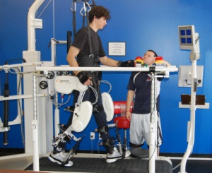
- Task-specific training with the aim of (re)acquiring a motor skill (with or without using robotic exoskeletons)
- Exercises that focus on regaining or sustaining control of the proximal muscles of the trunk, shoulder and pelvic girdle
- Exercises that aim to improve static and dynamic balance and proprioception as a component of postural control
- Stretching exercises that aim to improve range of movement.
- Adjuncts, such as treadmill training with or without partial body weight support, functional electrical stimulation of voluntary muscles and exergames that use computer technologies to provide an interactive environment which requires limb movement to react to on-screen gameplay (e.g. Wii, X Box).[6]

An important finding of a recent study[7] was that the level of challenge to balance was more important than the duration of exercise in producing Neurorehabilitation and neural repair. Cerebellar patients with ataxia can benefit from a home exercise program focused on balance training (with significant improvements after just six weeks). Highlighted was the importance of the 1. level of challenge to balance being of upmost importance 2. individualizing the program and offering continued training and progression if necessary to see effect retention.
Regeneration ? ![edit | edit source]
Current research is going on into the search for neurogenesis of the central nervous system. An exciting prospect. Some of the news is reported below.
- A current project is exploring the degree to which the brain can be repaired after neuron loss and identify factors that can stimulate stem cells to regenerate neurons.[8]
- A 2019 [9]) report shows compelling evidence that the mature central nervous system (CNS) harbors stem cell populations outside conventional neurogenic regions. It has been demonstrated that brain pericytes (PCs) in both mouse and human exhibit multipotency to differentiate into various neural lineages following cerebral ischemia. Importantly putative ischemia-induced multipotent stem cells are present in poststroke cerebellum and possess region-specific traits, suggesting a potential capacity to regenerate functional cerebellar neurons following ischemic stroke[9].
- Another 2019 study reported "Nerves in the central nervous system of adult mammals do not usually regenerate when injured. The granule cell, a nerve cell located in the cerebellum, is different. When its fibres, called parallel fibres, are cut, rapid regeneration ensues and junctions with other neurons called "synapses" are rebuilt. The precise mechanism for this was unclear"
Further Viewing[edit | edit source]
The below 4 minute video gives a great 3D view of the cerebellum and highlights it's similar structure to that of the cerebrum.
References[edit | edit source]
- ↑ 1.0 1.1 1.2 1.3 1.4 1.5 1.6 1.7 Jimsheleishvili S, Dididze M. Neuroanatomy, Cerebellum. InStatPearls [Internet] 2019 Mar 2. StatPearls Publishing.Available from:https://www.ncbi.nlm.nih.gov/books/NBK538167/ (last accessed 19.1.2020)
- ↑ 2.0 2.1 2.2 2.3 Teach me anatomy. The Cerebellum Available from: https://teachmeanatomy.info/neuroanatomy/structures/cerebellum/ (last accessed 20.1.2020)
- ↑ 3.0 3.1 3.2 Lumenlearning. Cerebellum Available from:https://courses.lumenlearning.com/boundless-ap/chapter/the-cerebellum/ (last accessed 20.1.2020)
- ↑ Soton brain hub Cerebellar disease symptoms Available from:https://www.youtube.com/watch?v=fMMcMPI68V0&app=desktop (last accessed 20.1.2020)
- ↑ Bastian AJ. Moving, sensing and learning with cerebellar damage. Current opinion in neurobiology. 2011 Aug 1;21(4):596-601.Available from:https://www.ncbi.nlm.nih.gov/pmc/articles/PMC3177958/ (last accessed 20.1.2020)
- ↑ Hartley H, Cassidy E, Bunn L, Kumar R, Pizer B, Lane S, Carter B. Exercise and Physical Therapy Interventions for Children with Ataxia: a systematic review. The Cerebellum. 2019 Aug 7:1-8.Available from:https://www.ncbi.nlm.nih.gov/pmc/articles/PMC6761087/ (last accessed 20.1.2020)
- ↑ Keller JL, Bastian AJ. A home balance exercise program improves walking in people with cerebellar ataxia. Neurorehabilitation and neural repair. 2014 Oct;28(8):770-8. Available from:https://www.ncbi.nlm.nih.gov/pmc/articles/PMC4133325/ (last accessed 20.1.2020)
- ↑ Sloan Kettering Inst. Cerebellum development and regeneration Available from: https://www.mskcc.org/research/ski/labs/alexandra-joyner/cerebellum-development-and-regeneration (last accessed 21.1.2020)
- ↑ 9.0 9.1 Beppu M, Nakagomi T, Takagi T, Nakano-Doi A, Sakuma R, Kuramoto Y, Tatebayashi K, Matsuyama T, Yoshimura S. Isolation and Characterization of Cerebellum-Derived Stem Cells in Poststroke Human Brain. Stem cells and development. 2019 Apr 15;28(8):528-42. Available from:https://www.liebertpub.com/doi/abs/10.1089/scd.2018.0232?journalCode=scd (last accessed 21.1.2020)
- ↑ Neuromatic The Cerebellum AVAILABLE FROM:https://www.youtube.com/watch?v=gqwCIXD0218&app=desktop (last accessed 21.1.2020)

