Basic Foot and Ankle Anatomy - Neural and Vascular
Original Editor - Ewa Jaraczewska
Top Contributors - Ewa Jaraczewska, Wanda van Niekerk, Jess Bell, Kim Jackson, Lucinda hampton and Olajumoke Ogunleye
Description[edit | edit source]
Identification of compression syndromes requires a very good understanding of the anatomy of the nerves and vessels of the foot and ankle. Morton's neuroma, Baxter neuropathy or jogger's heel (Plantar Fasciitis) syndromes are common pathologies requiring a physiotherapist's intervention.
Neural[edit | edit source]
Ankle and Foot[edit | edit source]
Tibial Nerve[edit | edit source]
The Tibial nerve originates at L5, S1 and S2 levels together with the common peroneal (fibular ) nerve.
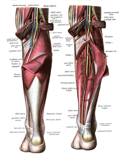
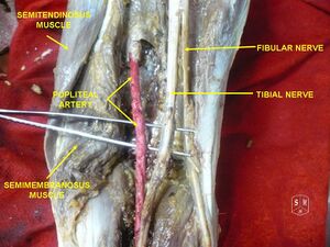
Distribution: popliteal fossa→between two heads of the gastrocnemius→medial border of the Achilles tendon→inferior and posterior to the medial malleolus→separates into calcaneal nerve brunches and medial and lateral plantar nerve→sole of the foot.
- Motor fibres: gastrocnemius, soleus, tibialis posterior, flexor digitorum longus, and flexor hallucis longus.
- Sensory fibres: occasionally supplies the area typically innervated by the deep peroneal nerve.[1]
Plantar Nerve[edit | edit source]
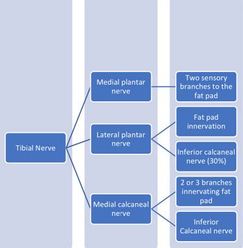
The plantar nerve originates from the tibial nerve at the medial malleolus. The nerve divides into two branches: medial and lateral. It provides motor and sensory innervation to the foot muscles.
- Medial plantar nerve
- Motor fibres: abductor hallucis, flexor digitorum brevis, and flexor hallucis brevis muscles.
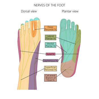
- Sensory fibres: sole innervation, 1st, 2nd and 3rd toes, often 4th toe.
- Lateral plantar nerve
- Motor fibres: abductor and flexor digiti minimi, the adductor hallucis, and the interossei muscles.
- Sensory fibres: sole innervation, 5th toe, occasionally 4th toe.
Calcaneal Nerve[edit | edit source]
The calcaneal nerve is a terminal branch of the tibial nerve.
- Medial calcaneal nerve
- Motor fibres: abductor hallucis
- Sensory fibres: medial border of the heel.
- Inferior calcaneal nerve (Baxter's nerve): originates from lateral plantar nerve (30%) and medial calcaneal nerve (70%).[2]
- Motor fibres: flexor digitorum brevis, quadratus plantae, abductor digiti minimi.
- Sensory fibres: plantar ligament, calcaneal periosteum.
Common Fibular (Peroneal) Nerve[edit | edit source]
The common fibular nerve originates at nerve roots L4, S1, and S2 together with the tibialis nerve.
Distribution: popliteal fossa→medial border of the biceps femoris→lateral head of the gastrocnemius→neck of the fibula→between the attachments of the fibularis longus muscle→divides into superficial and deep fibular nerves.
- Superficial
- Motor fibres: fibularis longus and brevis
- Sensory fibres: skin of the anterolateral leg, and dorsum of the foot (except the skin between the first and second toes).
- Deep: occasionally becomes the pure sensory nerve[1]
- Motor fibres: tibialis anterior, extensor digitorum longus, extensor hallucis longus, extensor digitorum brevis (rarely innervated by the tibial nerve).[1]
- Sensory fibres: lateral part of the ankle and foot regions, the skin between the first and second toes.
Sural Nerve[edit | edit source]
The Sural nerve originates from the tibial nerve and cutaneous branches of the common fibular nerve. It is divided into the sural communication nerve and lateral sural cutaneous nerve.
- Sensory fibres: posterior aspect of the distal leg and lateral aspect of the foot.
Vascular[edit | edit source]
Ankle and Foot[edit | edit source]
Popliteal Artery[edit | edit source]
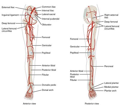
- Distribution: femoral artery→popliteal fossa→between the gastrocnemius and popliteal muscles in the posterior compartment of the leg→between the two heads of the gastrocnemius→divides into anterior and posterior tibial arteries.
- Supply: superficial posterior compartment including gastrocnemius, soleus and plantaris muscles.
Tibial Artery[edit | edit source]
Anterior tibial artery
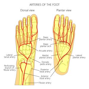
- Distribution: popliteal fossa→lower border of the popliteus muscle→posterior compartment of the leg→between heads of the posterior tibialis muscle→proximal part of the interosseous membrane→medial to the fibular neck→anterior compartment of the leg→anterior aspect of the interosseous membrane →anterior surface of the tibia→dorsalis pedis artery
- Supply: proximal tibiofibular joint, knee joint, ankle joint, muscles and skin of the anterior compartment of the leg.
Posterior tibial artery
- Distribution: terminal branch of the popliteal artery at the lower margin of the popliteus muscle→posterior compartment of the leg→inferior to the medial malleolus→tubercle of calcaneus→lateral and medial plantar arteries.
- Supply: soleus muscle, popliteus, flexor hallucis longus, flexor digitorum longus and tibialis posterior.
Fibular (Peroneal) Artery[edit | edit source]
- Distribution: posterior tibial artery, distal to the popliteus muscle→medial side of the fibula→inferior tibiofibular syndesmosis→calcaneal branches.
- Supply: popliteus, soleus, tibialis posterior, and flexor hallucis longus muscles.
Plantar Artery[edit | edit source]
- Medial and lateral plantar arteries form a plantar arch extending from the 1st to the 5th metatarsal. It joins a deep plantar branch of the dorsalis pedis.
- The medial plantar artery supplies abductor hallucis muscle and flexor digitorum brevis muscle.
- The lateral plantar artery supplies plantar aponeurosis between flexor digitorum brevis and abductor digiti minimi muscles.
Sural Artery[edit | edit source]
- The sural artery originates from each side of the popliteal artery. It has two branches: lateral and medial.
- Supply: gastrocnemius muscle, soleus muscle and plantaris muscle.
Clinical relevance[edit | edit source]
- Inferior calcaneal nerve entrapment (Baxter neuropathy) presents with heel pain and paraesthesia with the abductor digiti minimi muscle weakness.[4]
- When the common fibular nerve is damaged, the patient may not be able to dorsiflex and evert the foot and extend the digits.
- Sural artery flap is used for the reconstruction of soft tissue defects around the lower third of the leg, dorsum of the foot, malleoli and hindfoot.[5]
Resources[edit | edit source]
Anatomy of the Ankle Ligaments: A Pictorial Essay - In this pictorial essay, the ligaments around the ankle are grouped, depending on their anatomic orientation, and each of the ankle ligaments is discussed in detail.
References[edit | edit source]
- ↑ 1.0 1.1 1.2 Yamashita M, Mezaki T, Yamamoto T. "All tibial foot" with sensory crossover innervation between the tibial and deep peroneal nerves. J Neurol Neurosurg Psychiatry. 1998 Nov;65(5):798-9.
- ↑ Louisia S, Masquelet AC. The medial and inferior calcaneal nerves: an anatomic study. Surg Radiol Anat. 1999;21(3):169-73.
- ↑ Anatomy Zone. Foot Arteries - 3D Anatomy Tutorial. 2015 Available from: https://www.youtube.com/watch?v=wLcy4JXgE18 [last accessed 24/08/2022]
- ↑ Bauones, S., Feger, J. Baxter neuropathy. Reference article, Radiopaedia.org. (accessed 25/12/ 2021) https://doi.org/10.53347/rID-25994
- ↑ Hashmi DPM, Musaddiq A, Ali DM, Hashmi A, Zahid DM, Nawaz DZ. Long-Term Clinical and Functional Outcomes of Distally Based Sural Artery Flap: A Retrospective Case Series. JPRAS Open. 2021 Jul 31;30:61-73.
