Avulsion Fractures of the Ankle
Original Editors - Niels Verbeeck
Top Contributors - Niels Verbeeck, Kim Jackson, Admin, Shaimaa Eldib, Pierreux Maarten, Scott Cornish, Uchechukwu Chukwuemeka, Vidya Acharya, Rachael Lowe, Jan Alderweireldt, Claire Knott, Wanda van Niekerk, Lucinda hampton and 127.0.0.1
Definition/Description:[edit | edit source]
An avulsion fracture is where a fragment of bone is pulled away at the ligamentous or tendinous attachment. It can be caused by a traumatic traction (repetitive long-term or a single high impact traumatic traction) of the ligament or tendon.This occurs as tendons can bear more load than the bone.[1][2] It can occur at numerous sites in the body, but some areas are more sensitive to these types of fractures than others, such as at the ankle which mostly occur at the lateral aspect of medial malleolus or in the foot where avulsion fractures are commonly at the base of the fifth metatarsal, but also at the talus and calcaneus. A twisting injury to the ankle and foot may cause an avulsion fracture at any of these locations.
Clinically Relevant Anatomy :[edit | edit source]
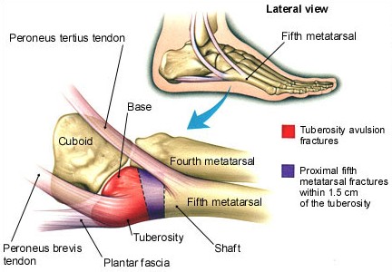
Taking the example of avulsion fractures at the 5th metatarsal; The metatarsal is divided into 3 parts: the tuberosity, the metaphysis and the head. Peroneus brevis attaches at the lateral side of the tuberosity (red area in figure 1), with peroneus tertius attaching at the dorsal side of the most proximal compartment of the metaphysis. Due to the high traction forces by these structures, tuberosity avulsion fractures commonly occur in an inversion injury (between the red and purple areas)
figure1: source: http://www.med-info.nl/images/images_trauma/Trauma_voet_MT5_Jones_groot.jpg
Characteristics/Clinical Presentation:[edit | edit source]
The characteristics of an avulsion fracture differ from those of a ligament rupture. Unlike non-operative treatment of a lateral ligament rupture, non-operative treatment of avulsion fractures do not yield satisfactory results. Symptoms of an ankle avulsion fracture are very similar to an ankle sprain and it is very difficult to diiferentiate without an X-ray or an MRI scan.
Pain is usually felt in the ankle immediately post injury with an immediate onset of swelling. Bruising may develop and the patient will have difficulty walking or weight bearing on the ankle. If an avulsion fracture is present, there will be immediate pain over the outside aspect of the foot. and associated with significant swelling and localised tenderness over 5th metatarsal. History of the injury will be similar to that of an ankle sprain (plantarflexor inversion). [3][4][5]
Differential Diagnosis:[edit | edit source]
- A Jones fracture occurs as a result of a stress fracture to the 5th metatarsal, due to repetitive loading of the outside part of the foot from the patient’s underlying foot pattern or lower extremity alignment. Unlike a Dancer’s fracture a Jones fracture may not heal and often requires surgery.[6]
- Stress fractures
- Mid-shaft fractures
Diagnostic Procedures:[edit | edit source]
An X-ray may be ordered by the surgeon. Avulsion fractures can sometimes be overlooked or where an injury to the 5th metatarsal occurs together with an ankle sprain. Other imaging methods are recommended such as, MRI, CT or scintigrams. [2] [7] [8]
Outcome Measures:[edit | edit source]
- Olerud ankle score
xamination:[edit | edit source]
A history of the injury is taken, such as mechanism, immediate pain levels and swelling. The Ottawa Ankle Rules can be used to localise the exact area of pain. Palpation may also be useful.
Medical Management:[edit | edit source]
An avulsion fracture of the base of the 5th metatarsal is usually treated conservatively. If the bone is not displaced, a walking boot or a walking cast can be used, which will remain in situ for 4 to 6 weeks. Surgery is only recommended where the bone is displaced from its normal position or where more than 30% of the cubometatarsal joint is involved. The bone will be removed or fixed with osteosynthesis material. Crutches may be used to avoid weight bearing on the injured foot. [9]
Physical Therapy Management:[edit | edit source]
Avulsion fractures are often treated as ankle sprains, with the dysfunctional movement and impairments treated alongside the fracture, so it is important to individualise the treatment plan. [8] Level of evidence: 2B
An inappropriately managed avulsion fractures can lead to significant, long-term functional disability. Most fractures heal well, but following a strict immobilisation period normal arthrokinematics, strength of the lower extremity muscles, proprioception and functional movement for chosen sport/activities need to be regained.
Rehabilitation following an avulsion fracture consists of 3 phases; the acute, the recovery and the functional phase:
- Acute phase: can begin at 2 weeks post-operatively. This phase can include passive range of motion exercises and crytherapy and is based on the reduction of pain, inflammation and oedema, while keeping muscle atrophy of the lower limb to a minimum.
- Recovery phase: begins once the goals of the acute phase have been met.
This phase can be further divided into 3 stages:
Weeks 0-6: active ROM exercises for the toes and the MPT joints, strengthening exercises for the ankle and foot are still premature however. In week 2, isometric exercises for the dorsiflexors, plantarflexors, invertors and evertors of the foot, along with active ankle ROM movements can be started.
Weeks 6-8: active and passive ROM exercises for the ankle and the subtalar joint with Isometric and isotonic exercises. Exercises for proprioception and proximal strength and control.
At 8-12 weeks: strengthening exercises for the dorsiflexors, plantarflexors, invertors, evertors, long flexors and extensors of the toes are recommended. Full weight bearing exercises are also permitted.[10][11] Level of evidence: 5 (10) en 4 (11) - Functional phase: is started at 6 to 8 weeks post-operatively. This phase involves ongoing strength and conditioning of the the lower limb, increasing neuromuscular control and utilising sport/activity specific training. [12] Level of evidence: 5
References:[edit | edit source]
- ↑ http://orthopedics.about.com/od/brokenbones/a/avulsion.htm
- ↑ 2.0 2.1 http://www.foothealthfacts.org/footankleinfo/fifth-metatarsal_fractures.htm
- ↑ Fracture Dislocations of the Tarsometatarsal Joints: End Results Correlated with Pathology and Treatment Level of evidence: 2A
- ↑ Haraguchi N, Toga H, Shiba N, Kato F, Avulsion fracture of the lateral ankle ligament complex in severe inversion injury: incidence and clinical outcome, Am J Sports Med. 2007 Jul;35(7):1144-52
- ↑ http://www.epainassist.com/sports-injuries/ankle-injuries/ankle-avulsion-fracture-symptoms-causes-treatment
- ↑ http://www.epainassist.com/sports-injuries/ankle-injuries/ankle-avulsion-fracture-symptoms-causes-treatment
- ↑ Duke G. Pao, Theodore E. Keats, Robert G. Dussault, Avulsion Fracture of the Base of the Fifth Metatarsal Not Seen on Conventional Radiography of the Foot: The Need for an Additional Projection, AJR 2000;175:549–552
- ↑ 8.0 8.1 Level of Evidence: 2B Haraguchi N, Toga H, Shiba N, Kato F, Avulsion fracture of the lateral ankle ligament complex in severe inversion injury: incidence and clinical outcome, Am J Sports Med. 2007 Jul;35(7):1144-52. Level of Evidence: 2B
- ↑ E.W. Zwitser *, R.S. Breederveld, Fractures of the fifth metatarsal; diagnosis and treatment, Injury, Int. J. Care Injured 41 (2010) 555–562
- ↑ Essentials of Orthopaedics for Physiotherapist, Ebnezar. Level of Evidence: 5
- ↑ Fracture Dislocations of the Tarsometatarsal Joints: End Results Correlated with Pathology and Treatment. Level of Evidence: 4
- ↑ http://www.podiatrytoday.com/article/6565 Level of Evidence: 5

