File:Tendinopathy of the Shoulder.png
Tendinopathy_of_the_Shoulder.png (570 × 488 pixels, file size: 186 KB, MIME type: image/png)
Summary[edit | edit source]
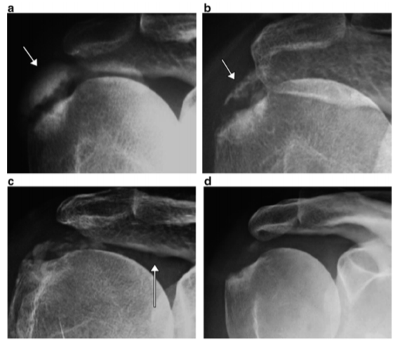
Fig. 6.16 X-ray images of a calcified tendinopathy of the shoulder ( a ) before ESWT, ( b ) and ( c ) 30 days after receiving a single session of 4000 shock waves (EFD = 0.35 mJ/mm 2
) with a Duluth
SD1 electromagnetic shock wave source (Storz Medical AG, Tägerwilen, Switzerland) and ( d ) 60 days after therapy. The arrow in ( a ) shows the extension of the calcifi cations. Small portions of calcium salts migrating medially can be seen ( arrow ) in the image ( c ). (Courtesy of L. Guiloff and M. Brañes)
Licensing[edit | edit source]
This file is copyrighted and not published under a free license, and the uploader asserts that the usage of this file is fair use.
File history
Click on a date/time to view the file as it appeared at that time.
| Date/Time | Thumbnail | Dimensions | User | Comment | |
|---|---|---|---|---|---|
| current | 13:09, 28 September 2020 | | 570 × 488 (186 KB) | Ayushi Tomer (talk | contribs) | Fig. 6.16 X-ray images of a calcified tendinopathy of the shoulder ( a ) before ESWT, ( b ) and ( c ) 30 days after receiving a single session of 4000 shock waves (EFD = 0.35 mJ/mm 2 ) with a Duluth SD1 electromagnetic shock wave source (Storz Medical... |
You cannot overwrite this file.
File usage
The following page uses this file:

