File:SIJ articular surfaces.jpg
SIJ_articular_surfaces.jpg (369 × 574 pixels, file size: 64 KB, MIME type: image/jpeg)
Summary[edit | edit source]
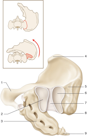
Fig. 1. Anatomy of the sacro-iliac joint: anterior view. 1. Body of the pubic bone. 2. Auricular surface of the sacrum. 3. Sacrum. 4. Antero-superior iliac spine. 5. Iliac tuberosity. 6. Auricular surface of the ilium. 7. Anterior sacro-iliac ligament. 8. Postero-superior iliac spine. 9. Coccyx. © Cyrille Martinet.
Open access from: Le Huec JC, Tsoupras A, Leglise A, Heraudet P, Celarier G, Sturresson B. The sacro-iliac joint: a potentially painful enigma. Update on the diagnosis and treatment of pain from micro-trauma. Orthopaedics & Traumatology: Surgery & Research. 2019 Feb 1;105(1):S31-42.
Licensing[edit | edit source]
This file is copyrighted and not published under a free license, and the uploader asserts that the usage of this file is fair use.
File history
Click on a date/time to view the file as it appeared at that time.
| Date/Time | Thumbnail | Dimensions | User | Comment | |
|---|---|---|---|---|---|
| current | 11:34, 22 January 2022 | | 369 × 574 (64 KB) | Abbey Wright (talk | contribs) | Fig. 1. Anatomy of the sacro-iliac joint: anterior view. 1. Body of the pubic bone. 2. Auricular surface of the sacrum. 3. Sacrum. 4. Antero-superior iliac spine. 5. Iliac tuberosity. 6. Auricular surface of the ilium. 7. Anterior sacro-iliac ligament. 8. Postero-superior iliac spine. 9. Coccyx. © Cyrille Martinet. Open access from: Le Huec JC, Tsoupras A, Leglise A, Heraudet P, Celarier G, Sturresson B. The sacro-iliac joint: a potentially painful enigma. Update on the diagnosis and treatme... |
You cannot overwrite this file.
File usage
The following page uses this file:

