File:Otoconia within the posterior canal endolymphatic duct.png
Otoconia_within_the_posterior_canal_endolymphatic_duct.png (716 × 538 pixels, file size: 246 KB, MIME type: image/png)
Summary[edit | edit source]
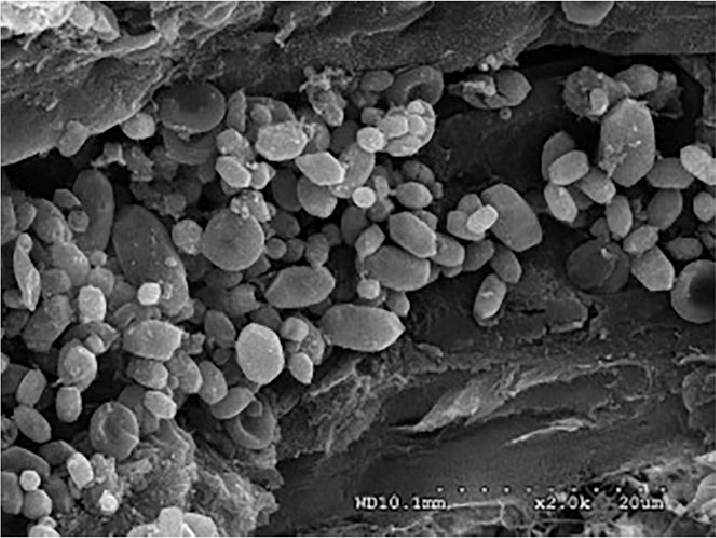
Benign paroxysmal positional vertigo - Scientific Figure on ResearchGate. Available from: https://www.researchgate.net/figure/Scanning-electron-micrograph-image-of-otoconia-within-the-posterior-canal-endolymphatic_fig2_329648423 [accessed 22 Jun, 2021]
Licensing[edit | edit source]
This file is licensed under the Creative Commons Attribution 4.0 Unported license.
You are free to:
- Share - copy and redistribute the material in any medium or format
- Adapt - remix, transform, and build upon the material for any purpose, even commercially.
The licensor cannot revoke these freedoms as long as you follow the license terms.
Under the following conditions:
- Attribution – You must give appropriate credit, provide a link to the license, and indicate if changes were made. You may do so in any reasonable manner, but not in any way that suggests the licensor endorses you or your use.
No additional restrictions — You may not apply legal terms or technological measures that legally restrict others from doing anything the license permits.
File history
Click on a date/time to view the file as it appeared at that time.
| Date/Time | Thumbnail | Dimensions | User | Comment | |
|---|---|---|---|---|---|
| current | 00:17, 23 June 2021 | | 716 × 538 (246 KB) | Jess Bell (talk | contribs) | Benign paroxysmal positional vertigo - Scientific Figure on ResearchGate. Available from: https://www.researchgate.net/figure/Scanning-electron-micrograph-image-of-otoconia-within-the-posterior-canal-endolymphatic_fig2_329648423 [accessed 22 Jun, 2021] |
You cannot overwrite this file.
File usage
The following page uses this file:

