File:HIV-budding-Color.jpg
Original file (2,967 × 1,971 pixels, file size: 3.92 MB, MIME type: image/jpeg)
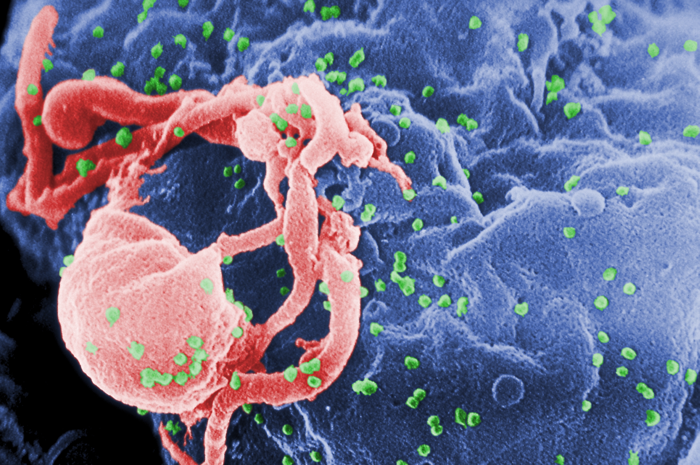
Summary[edit | edit source]
Scanning electron micrograph of HIV-1 budding (in green) from cultured lymphocyte. This image has been colored to highlight important features; see PHIL 1197 for original black and white view of this image. Multiple round bumps on cell surface represent sites of assembly and budding of virions. https://commons.wikimedia.org/wiki/File:HIV-budding-Color.jpg
Photo Credit: C. GoldsmithContent Providers: CDC/ C. Goldsmith, P. Feorino, E. L. Palmer, W. R. McManus [Public domain]
Licensing[edit | edit source]
This file is a work of an employee of the US federal government, taken or made during the course of the person's official duties. As a work of the U.S. federal government, the image is in the public domain.
File history
Click on a date/time to view the file as it appeared at that time.
| Date/Time | Thumbnail | Dimensions | User | Comment | |
|---|---|---|---|---|---|
| current | 21:06, 23 November 2019 | | 2,967 × 1,971 (3.92 MB) | Kim Jackson (talk | contribs) | Scanning electron micrograph of HIV-1 budding (in green) from cultured lymphocyte. This image has been colored to highlight important features; see PHIL 1197 for original black and white view of this image. Multiple round bumps on cell surface represen... |
You cannot overwrite this file.
File usage
The following 2 pages use this file:

