File:Facial nerve schwannoma.png
No higher resolution available.
Facial_nerve_schwannoma.png (512 × 514 pixels, file size: 198 KB, MIME type: image/png)
Summary[edit | edit source]
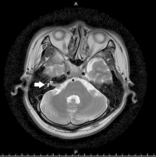
Author: Chen MC, Tseng TM, Hung SH, Chen PY Description: Contrast-enhanced T2-weighted magnetic resonance imaging reveals an intracanalicular tumor in the region of the cerebellopontine angle (shown by arrow). Creation time 2014
Licensing[edit | edit source]
This file is licensed under the Creative Commons Attribution-Share Alike 3.0 Unported license.
You are free:
- to share
- to copy, distribute and transmit the work to remix
- to adapt the work
Under the following conditions:
- attribution – You must attribute the work in the manner specified by the author or licensor (but not in any way that suggests that they endorse you or your use of the work).
- share alike – If you alter, transform, or build upon this work, you may distribute the resulting work only under the same or similar license to this one.
File history
Click on a date/time to view the file as it appeared at that time.
| Date/Time | Thumbnail | Dimensions | User | Comment | |
|---|---|---|---|---|---|
| current | 21:38, 28 November 2019 | | 512 × 514 (198 KB) | Wendy Walker (talk | contribs) | Author: Chen MC, Tseng TM, Hung SH, Chen PY Description: Contrast-enhanced T2-weighted magnetic resonance imaging reveals an intracanalicular tumor in the region of the cerebellopontine angle (shown by arrow). Creation time 2014 |
You cannot overwrite this file.
File usage
The following page uses this file:

