Tectorial membrane
Original Editor - Rachael Lowe
Top Contributors - Rachael Lowe, Priyanka Chugh, Kim Jackson and Evan Thomas
Description[edit | edit source]
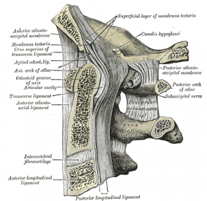
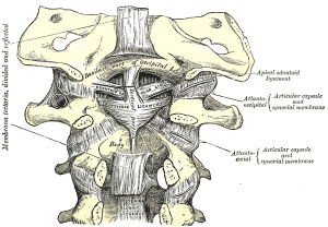
Strong broad structure which covers the Odontoid process and it's ligaments.
Continuous with the Posterior longitudinal ligament and found on the internal surface of the vertebral canal.
Attachments[edit | edit source]
Arises from the posterior surface of the body of the Axis and, expanding as it ascends, is attached to the basilar groove of the occipital bone, in front of the foramen magnum.
Its anterior surface is in relation with the Transverse ligament of the atlas and its posterior surface with the dura mater. As it enters the cranial cavity it becomes continuous with the dura mater.
Function[edit | edit source]
Contributes to the stability of the upper cervical spine. Limits Flexion (C0/C1 and C1/C2) and rotation (C0/C1)[1].
Histology[edit | edit source]
The tectorial membrane (TM) is an extracellular connective tissue that covers the mechanically-sensitive hair bundles of the sensory receptor cells in the inner ear. It occupies a strategic position, playing a key role in transforming sound to mechanical stimulation. The TM is synthesized by interdental cells (not shown) and is composed mainly by water (97%). The remaining solid fraction (protein and carbohydrate) forms a matrix that contains ionizable charge groups, attracting mobile counterions from the surrounding fluid. In the spiral canal, the Reissner's membrane The outer surface of the TM is covered by a covering net (left) of amorphous glycoprotein substance that continues over its border and fixes the TM to the Hensen's cells. The bulk of the TM has a striated appearance as it contains radial microfibrils 9-11 nm thick, embedded in a homogeneous glycosaminoglycan containing substrate. Charged glycoconjugates maybe responsible for ion sequestering within the TM. From a dark nonstriated band on the undersurface of the TM, called Hensen's stripe, fine gelatinous trabeculae extend to fix the TM to the border cells, near the inner hair cells.[2]
Examination[edit | edit source]
Recent Related Research (from Pubmed)[edit | edit source]
Failed to load RSS feed from http://www.ncbi.nlm.nih.gov/entrez/eutils/erss.cgi?rss_guid=1vsc_5ltFDWrFZny0uTaD-pGjWEAtnNS9oEyE4pVZB0DHMVMQA|charset=UTF-8|short|max=10: Error parsing XML for RSS
References[edit | edit source]
- ↑ T. Oda, M.M. Panjabi, J.J. Crisco, H.U. Bueff, D. Grob, J. Dvorak. Role of tectorial membrane in the stability of the upper cervical spine. Clinical Biomechanics, Volume 7, Issue 4, November 1992, Pages 201–207
- ↑ http://147.162.36.50/cochlea/cochleapages/anatomy/tect.htm


