Muscles of Respiration: Difference between revisions
Evan Thomas (talk | contribs) mNo edit summary |
Evan Thomas (talk | contribs) mNo edit summary |
||
| Line 14: | Line 14: | ||
== Accessory Muscles == | == Accessory Muscles == | ||
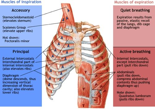
The accessory inspiratory muscles are the | The accessory inspiratory muscles are the sternocleidomastoid, the scalenus anterior, medius and posterior, the pectoralis major and minor, the inferior fibres of serratus anterior and latissimus dorsi, the serratus posterior anterior may help in inspiration also the iliocostalis cervicis<ref name="anatomy">http://voiceandalexandertechnique.eu/voice-anatomy/pharynx-and-larynx/muscles-involved-in-voice-production/muscles-of-respiration.html</ref>. | ||
The accessory expiratory muscles are the abdominal muscles: rectus, abdominis, external oblique, internal oblique and transversus abdominis. And in the thoracolumbar region the lowest fibres of iliocostalis and longissimus, the serratus posterior inferior and quadratus lumborum. | The accessory expiratory muscles are the abdominal muscles: rectus, abdominis, external oblique, internal oblique and transversus abdominis. And in the thoracolumbar region the lowest fibres of iliocostalis and longissimus, the serratus posterior inferior and quadratus lumborum. | ||
<br> | <br> | ||
| Line 24: | Line 22: | ||
[[Image:949 937 muscles-of-respiration.jpg]] | [[Image:949 937 muscles-of-respiration.jpg]] | ||
<br> | |||
<br> | |||
== Innervation of respiratory muscles:<ref name="snell">Snell's Clinical Anatomy http://teachinganatomy.blogspot.com/2013/07/respiratorymuscles.html</ref> == | == Innervation of respiratory muscles:<ref name="snell">Snell's Clinical Anatomy http://teachinganatomy.blogspot.com/2013/07/respiratorymuscles.html</ref> == | ||
Revision as of 18:13, 5 April 2017
Original Editors - Rachael Lowe
Top Contributors - Khloud Shreif, Andeela Hafeez, Candace Goh, Vidya Acharya, Rachael Lowe, George Prudden, Kim Jackson, Admin, Tomer Yona, Lucinda hampton, Evan Thomas, WikiSysop, Joao Costa, Lenny Vasanthan T, Sai Kripa, 127.0.0.1 and Rishika Babburu -
Introduction[edit | edit source]
The breathing pump muscles are a complex arrangement that form a semirigid bellows around the lungs. Essentially, all muscles that attach to the rib cage have the potential to generate a breathing action. Muscles that expand the thoracic cavity are inspiratory muscles and induce inhalation, while those that compress the thoracic cavity are expiratory and induce exhalation. These muscles possess exactly the same basic structure as all other skeletal muscles, and they work in concert to expand or compress the thoracic cavity.[1]
Primary Muscles[edit | edit source]
Accessory Muscles[edit | edit source]
The accessory inspiratory muscles are the sternocleidomastoid, the scalenus anterior, medius and posterior, the pectoralis major and minor, the inferior fibres of serratus anterior and latissimus dorsi, the serratus posterior anterior may help in inspiration also the iliocostalis cervicis[2].
The accessory expiratory muscles are the abdominal muscles: rectus, abdominis, external oblique, internal oblique and transversus abdominis. And in the thoracolumbar region the lowest fibres of iliocostalis and longissimus, the serratus posterior inferior and quadratus lumborum.
Innervation of respiratory muscles:[3][edit | edit source]
Attachment of Diaphragm:[3][edit | edit source]
Origin: Xiphoid process (posterior surface), lower six ribs and their costal ccartilage (inner surface) and upper three lumbar vertebra as right crus and upper two lumbar vertebra as left crus.
Insertion: central tendon
Nerve Supply: Motor nerve supply by Phrenic nerve (C3 C4 C5) and sensory supply by phrenic nerve to centrarl tendon and lower 6 or 7 intercostal nerve to peripheral parts.
Intercostal muscles:[3][edit | edit source]
They are three types: External intercostal muscles, internal intercostal muscles and innermost intercostal muscles.
External intercostal muscles:
Origin: inferior border of rib above and
Insertion: superior border of rib below
Internal intercostal muscles:
Origin: from the costal groove (lower part of inner surface of rib near the inferior border) of the rib above and
Insertion: upper border of rib below
Innermost intercostal muscles:
It is an incomplete muscle layer and crosses more than one intercostal space. These muscles assist in the function of external and internal intercostal muscles.
Origin: from the costal groove of the rib above and
Insertion: the superior border of rib below
Nerve supply: all the intercostal muscles are supplied by intercostal nerves
Recent Related Research (from Pubmed)[edit | edit source]
References[edit | edit source]
- ↑ Breathe Strong, Perform Better by Alison McConnell http://www.humankinetics.com/excerpts/excerpts/learn-the-anatomy-and-physiology-of-the-muscles-involved-in-breathing
- ↑ 2.0 2.1 http://voiceandalexandertechnique.eu/voice-anatomy/pharynx-and-larynx/muscles-involved-in-voice-production/muscles-of-respiration.html
- ↑ 3.0 3.1 3.2 Snell's Clinical Anatomy http://teachinganatomy.blogspot.com/2013/07/respiratorymuscles.html


