Muscle Spindles: Difference between revisions
Oyemi Sillo (talk | contribs) No edit summary |
Scott Buxton (talk | contribs) No edit summary |
||
| Line 32: | Line 32: | ||
<br> | <br> | ||
<br> | |||
<br> | |||
== Function == | == Function == | ||
| Line 40: | Line 40: | ||
Muscle spindles stimulate reflexively a muscle contraction to prevent overstretching and muscle fiber damage, this is known as the stretch or myotatic reflex <ref name="idiopathic" />. While stretching the muscle spindle, an impulse is immediately sent to the spinal cord and a response to contract the muscle is received, for protecting it from being pulled forcefully or beyond a normal range. It is a very quick impulse, because the impulse only has to go to the spinal cord and back <ref name="spasticity" />. The stretch reflex has two components. The static component lasts as long as the muscle is being stretched. The dynamic component lasts for only a moment in response to the initial sudden increase in muscle length. The purpose of muscle spindles and the stretch reflex is to protect your body from injury caused by overstretching and to maintain muscle tone <ref name="changes">Hospod V., Aimonetti J.-M., Roll J.-P., Ribot-Ciscar E. Changes in human muscle spindle sensitivity during proprioceptive attention task. The Journal of Neuroscience.2001;27:19:5172-5178. Level of evidence: A2</ref>. This is a process that inhibits the stretch reflex in antagonistic pairs of muscles. An impulse is sent from the stretched muscle spindle when the stretch reflex is activated, the motor neuron is split so that the signal to contract can be sent to the stretched muscle, while a signal to relax can be sent to the antagonist muscles <ref>Kakuda N., Nagaoka M. Dynamic response of human muscle spindle afferents to stretch during voluntary contraction. Journal of Physiology. 1998;513:2:621-628. Level of evidence: A2</ref>. | Muscle spindles stimulate reflexively a muscle contraction to prevent overstretching and muscle fiber damage, this is known as the stretch or myotatic reflex <ref name="idiopathic" />. While stretching the muscle spindle, an impulse is immediately sent to the spinal cord and a response to contract the muscle is received, for protecting it from being pulled forcefully or beyond a normal range. It is a very quick impulse, because the impulse only has to go to the spinal cord and back <ref name="spasticity" />. The stretch reflex has two components. The static component lasts as long as the muscle is being stretched. The dynamic component lasts for only a moment in response to the initial sudden increase in muscle length. The purpose of muscle spindles and the stretch reflex is to protect your body from injury caused by overstretching and to maintain muscle tone <ref name="changes">Hospod V., Aimonetti J.-M., Roll J.-P., Ribot-Ciscar E. Changes in human muscle spindle sensitivity during proprioceptive attention task. The Journal of Neuroscience.2001;27:19:5172-5178. Level of evidence: A2</ref>. This is a process that inhibits the stretch reflex in antagonistic pairs of muscles. An impulse is sent from the stretched muscle spindle when the stretch reflex is activated, the motor neuron is split so that the signal to contract can be sent to the stretched muscle, while a signal to relax can be sent to the antagonist muscles <ref>Kakuda N., Nagaoka M. Dynamic response of human muscle spindle afferents to stretch during voluntary contraction. Journal of Physiology. 1998;513:2:621-628. Level of evidence: A2</ref>. | ||
<br> | |||
== Resources == | == Resources == | ||
[http://www.slideshare.net/biotechvictor1950/physiology-of-muscle-contraction-17064052?qid=356470bf-d9b7-4e5a-8528-d8e88cf3ac0c&v=qf1&b=&from_search=7 The Physiology of Muscle Contraction] by St. Xavier's College | [http://www.slideshare.net/biotechvictor1950/physiology-of-muscle-contraction-17064052?qid=356470bf-d9b7-4e5a-8528-d8e88cf3ac0c&v=qf1&b=&from_search=7 The Physiology of Muscle Contraction] by St. Xavier's College | ||
== Recent Related Research (from Pubmed) == | == Recent Related Research (from Pubmed) == | ||
| Line 50: | Line 50: | ||
== References == | == References == | ||
<references /> | <references /> | ||
[[Category:Cerebral_Palsy]] | |||
Revision as of 19:48, 6 July 2016
Original Editor - Bo Hellinckx
Top Contributors - Tania Appelmans, Adam Vallely Farrell, Kirenga Bamurange Liliane, Lucinda hampton, Laura Ritchie, Vidya Acharya, Oyemi Sillo, Bo Hellinckx, Scott Buxton, Wanda van Niekerk, Cindy John-Chu, Ahmed M Diab, Rachael Lowe, Evan Thomas, WikiSysop and Kim Jackson
Definition[edit | edit source]
Muscle spindles are skeletal muscle sensory organs that contribute to fine motor control and provide axial and limb position information to the central nervous system. They are involved in the sensation of position and movement of the body, this is called proprioception [1][2].
Composition[edit | edit source]
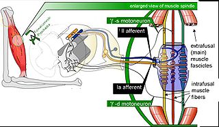
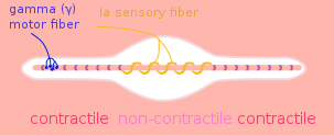
Muscle spindles are small sensory organs, with an elongated shape. A muscle spindle contains intrafusal fibers. Those are several small, specialized muscle fibers. Those fibers are oriented parallel to the regular, power-producing extrafusal muscle fibers. Intrafusal muscle fibers are at both ends connected to either tendinous ligaments or extrafusal fibers, namely contractile proteins [2]. So, intrafusal fibers are stretched or shortened correspondingly, when extrafusal fibres change length. The central part of the muscle spindle is covered with a capsule of connective tissue. The sensory dendrites of the muscle spindle afferent wrap the central region. The muscle spindle is stretched when the muscle lengthens increase, this opens mechanically-gated ion channels in the sensory dendrites. This leads to a receptor potential that triggers action potentials in the muscle spindle afferent [3][4]. There can be found two types of sensory endings in muscle spindles: the primary and secondary endings of spindles, they are located in the middle of the spindle. The primary endings respond to its speed and the size of a muscle length change. They belong to the fastest axons, because they are myelinated. They contribute both to movement and the sense of limb position. Secondary endings are only sensitive to length and not to velocity, so they contribute only to the sense of the position. These endings have smaller axons and thus slower conduction speed. Both endings in muscle spindles are very sensitive to low-amplitude changes in muscle length, especially if these changes occur at a high frequency. A spindle ending is located at the end of a neuron or an axon whose body is in the spinal ganglion [2][5].
Function[edit | edit source]
Muscle spindles stimulate reflexively a muscle contraction to prevent overstretching and muscle fiber damage, this is known as the stretch or myotatic reflex [1]. While stretching the muscle spindle, an impulse is immediately sent to the spinal cord and a response to contract the muscle is received, for protecting it from being pulled forcefully or beyond a normal range. It is a very quick impulse, because the impulse only has to go to the spinal cord and back [4]. The stretch reflex has two components. The static component lasts as long as the muscle is being stretched. The dynamic component lasts for only a moment in response to the initial sudden increase in muscle length. The purpose of muscle spindles and the stretch reflex is to protect your body from injury caused by overstretching and to maintain muscle tone [5]. This is a process that inhibits the stretch reflex in antagonistic pairs of muscles. An impulse is sent from the stretched muscle spindle when the stretch reflex is activated, the motor neuron is split so that the signal to contract can be sent to the stretched muscle, while a signal to relax can be sent to the antagonist muscles [6].
Resources[edit | edit source]
The Physiology of Muscle Contraction by St. Xavier's College
Recent Related Research (from Pubmed)[edit | edit source]
References[edit | edit source]
- ↑ 1.0 1.1 Grünewald R.A., Yoneda Y., Shipman J.M., Sagar H.J. Idiopathic focal dystonia: a disorder of muscle spindle afferent processing. Brain. 1997;120:2179-2185. Level of evidence: A2
- ↑ 2.0 2.1 2.2 Proske U., Candevia S.C. The kineasthetic senses. Journal of Physiology. 2009;17:4139-4146. Level of evidence: A1
- ↑ Soukup T., Thernell L.-E. Unusual intrafusal fibres in human muscle spindles. Physiol. Res. 1999;48:519-523. Level of evidence: B2
- ↑ 4.0 4.1 Mukherjee A., Chakravarty Ambar. Spasticity mechanisms for the clinician. Frontiers in Neurology. 2010;1:149:1-10. Level of evidence: A1
- ↑ 5.0 5.1 Hospod V., Aimonetti J.-M., Roll J.-P., Ribot-Ciscar E. Changes in human muscle spindle sensitivity during proprioceptive attention task. The Journal of Neuroscience.2001;27:19:5172-5178. Level of evidence: A2
- ↑ Kakuda N., Nagaoka M. Dynamic response of human muscle spindle afferents to stretch during voluntary contraction. Journal of Physiology. 1998;513:2:621-628. Level of evidence: A2


