Medial Plantar Nerve: Difference between revisions
No edit summary |
No edit summary |
||
| Line 9: | Line 9: | ||
[[Image:Gray833.png|thumb|right|250x300px]] | [[Image:Gray833.png|thumb|right|250x300px]] | ||
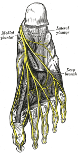
The medial plantar nerve is the larger one of the two terminal branches of the tibial nerve, it covers most of the sole of the foot and supply multiple muscles which functioning on the toes. | The medial plantar nerve is the larger one of the two terminal branches of the tibial nerve, it covers most of the sole of the foot and supply multiple muscles which functioning on the toes. | ||
== '''Anatomy <ref name="Clinical anatomy">Richard S. Snell,1992,Clinical Anatomy for Medical Students,fourth edition,little brown and company,Boston</ref>''' == | == '''Anatomy <ref name="Clinical anatomy">Richard S. Snell,1992,Clinical Anatomy for Medical Students,fourth edition,little brown and company,Boston</ref>''' == | ||
| Line 31: | Line 31: | ||
=== Movements produced: === | === Movements produced: === | ||
Flexion and abduction of the big toe (flexor hallucis brevis and abductor hallucis) | Flexion and abduction of the big toe (flexor hallucis brevis and abductor hallucis) | ||
Flexion of the toes (flexor digitorum brevis and the first lumbrical muscle) | Flexion of the toes (flexor digitorum brevis and the first lumbrical muscle) | ||
== Pathology/Injury == | == Pathology/Injury == | ||
'''Medial plantar nerve entrapment:''' | '''Medial plantar nerve entrapment:''' | ||
| Line 41: | Line 41: | ||
It is a compression of the nerve branches, where the nerve branches are compressed between bones, ligaments and other connective tissues causing a pain at the inner heel area. | It is a compression of the nerve branches, where the nerve branches are compressed between bones, ligaments and other connective tissues causing a pain at the inner heel area. | ||
Symptoms include almost constant pain whenever adding a pressure to the foot either by walking or sitting, just standing is often difficult.<ref name="Med.p">Medial and Lateral Plantar Nerve Entrapment, http://www.msdmanuals.com/home, (accessed 11 Jan 2017)</ref> | Symptoms include almost constant pain whenever adding a pressure to the foot either by walking or sitting, just standing is often difficult.<ref name="Med.p">Medial and Lateral Plantar Nerve Entrapment, http://www.msdmanuals.com/home, (accessed 11 Jan 2017)</ref> | ||
== Physiotherapy Assessment == | == Physiotherapy Assessment == | ||
| Line 47: | Line 47: | ||
=== Observation: === | === Observation: === | ||
Local observation for the sole of the foot is the first step of examination, notice any difference | Local observation for the sole of the foot is the first step of examination, notice any difference compared with the unaffected side, injury or incision, bruises, lump and the skin colour on the related area. | ||
*The atrophy muscle is a sign to indicate if there is an impairment of the nerve that innervates the affected muscle, but it is difficult to be | *The atrophy muscle is a sign to indicate if there is an impairment of the nerve that innervates the affected muscle, but it is difficult to be recognised with small muscles. | ||
=== Palpation: === | === Palpation: === | ||
| Line 57: | Line 57: | ||
=== Manual muscle test: === | === Manual muscle test: === | ||
Examine the strength of the muscles that Innervated by the medial plantar nerve, by resisting the movement of the big toe flexion/abduction and toes flexion. | |||
== Recent Related Research (from [http://www.ncbi.nlm.nih.gov/pubmed/ Pubmed]) == | == Recent Related Research (from [http://www.ncbi.nlm.nih.gov/pubmed/ Pubmed]) == | ||
| Line 66: | Line 66: | ||
References will automatically be added here, see [[Adding References|adding references tutorial]]. | References will automatically be added here, see [[Adding References|adding references tutorial]]. | ||
<references /></div> | |||
<references /> | |||
Revision as of 21:41, 11 January 2017
Original Editor - Your name will be added here if you created the original content for this page.
Lead Editors - Asma Alshehri, Kim Jackson, George Prudden, Shaniel Walters, Rachael Lowe, Tony Lowe, WikiSysop, Jaroslaw Pospiech and Wendy Snyders
Description
[edit | edit source]
The medial plantar nerve is the larger one of the two terminal branches of the tibial nerve, it covers most of the sole of the foot and supply multiple muscles which functioning on the toes.
Anatomy [1][edit | edit source]
General Course of Nerve:[edit | edit source]
It arises under the flexor retinaculum and runs forward deep to the abductor hallucis with the medial plantar artery on its medial side. It comes to lie in the interval between the abductor hallucis and the flexor digitorum brevis.
Branches:[edit | edit source]
Cutaneous branches: plantar digital nerves run to the sides of the medial three and the medial half of the fourth toe. The nerves extend onto the dorsumand supply the nail beds and the tips of the toes.
Muscular branches: it gives a branches to these four muscles, abductor hallucis, flexor digitorum brevis, the flexor hallucis brevis and the first lumbrical muscle.
Function[edit | edit source]
Innervates (sensory and motor):
[edit | edit source]
See the branches section above.
Movements produced: [edit | edit source]
Flexion and abduction of the big toe (flexor hallucis brevis and abductor hallucis)
Flexion of the toes (flexor digitorum brevis and the first lumbrical muscle)
Pathology/Injury[edit | edit source]
Medial plantar nerve entrapment:
It is a compression of the nerve branches, where the nerve branches are compressed between bones, ligaments and other connective tissues causing a pain at the inner heel area.
Symptoms include almost constant pain whenever adding a pressure to the foot either by walking or sitting, just standing is often difficult.[2]
Physiotherapy Assessment[edit | edit source]
Observation:[edit | edit source]
Local observation for the sole of the foot is the first step of examination, notice any difference compared with the unaffected side, injury or incision, bruises, lump and the skin colour on the related area.
- The atrophy muscle is a sign to indicate if there is an impairment of the nerve that innervates the affected muscle, but it is difficult to be recognised with small muscles.
Palpation:[edit | edit source]
You can assess the sensation of the areas supplied by the medial plantar nerve and palpate the related area to check any problems relating to the sensation (either hyper sensitivity or impaired sensation) and/or the tenderness degree.
Manual muscle test: [edit | edit source]
Examine the strength of the muscles that Innervated by the medial plantar nerve, by resisting the movement of the big toe flexion/abduction and toes flexion.
Recent Related Research (from Pubmed)[edit | edit source]
Extension:RSS -- Error: Not a valid URL: Feed goes here!!|charset=UTF-8|short|max=10
References[edit | edit source]
References will automatically be added here, see adding references tutorial.
- ↑ Richard S. Snell,1992,Clinical Anatomy for Medical Students,fourth edition,little brown and company,Boston
- ↑ Medial and Lateral Plantar Nerve Entrapment, http://www.msdmanuals.com/home, (accessed 11 Jan 2017)


