Bankart lesion
Original Editors
Top Contributors - Nikki Rommers, Estelle Hovaere, Lucinda hampton, Chrysolite Jyothi Kommu, Kim Jackson, Julie Schuermans, Admin, WikiSysop, Birgit Schauvliege, Wanda van Niekerk, 127.0.0.1, Elodie Baele, Naomi O'Reilly and Fasuba Ayobami
Search Strategy[edit | edit source]
- bankart lesion
- rehabilitation
- operative repair
- shoulder dislocation
- Bankart lesion treatment
- Bankart medical management
- Therapy bankart
- Bankart medical
- Open bankart
- Measurements shoulder
- WOSI
- ISIS
Definition/Description[edit | edit source]
A Bankart lesion[1] is a lesion of the anterior part of the glenoid labrum of the shoulder. This injury is caused by repeated anterior shoulder subluxations. The dislocation of the shoulder joint (anterior) can damage the connective tissue ring around the glenoid labrum. It can also bring damage to the connection between the labrum and capsule. Usually it has to do with none or poorly construction of the medial glenohumeral ligament. This injury is common for athletes that practice volleyball, tennis, handball, people who do overhead activities,…
Clinically Relevant Anatomy[edit | edit source]
The Bankart lesion is an injury of the glenohumeral joint. This is a ball-and-socket joint binds the scapular and the humerus. Parts of the joint are the labrum, a fibrocartilaginous structure around the glenoid, the capsule and ligaments and supporting muscle tendons.[2][3]
Epidemiology /Etiology[edit | edit source]
The shoulder is designed for its mobility, with stability being sacrificed to achieve this mobility. Due to poor osseous congruency and capsular laxity, the glenohumeral joint is very unstable, which makes it the most frequently dislocated joint in the human body. It relies on dynamic stabilizers and the neuromuscular system for its stability. Anterior instability is the most common traumatic type of instability, representing approximately 95% of all shoulder instabilities. [4] Glenohumeral dislocations are mainly caused by an abduction, extension and external rotation movement. [2][4][3]
In many cases of anterior dislocation patients have a Bankart lesion.
A reverse Bankart lesion can occurs in case of a posterior dislocation.
Characteristics/Clinical [edit | edit source]
We can distinguish to types of Bankart lesions: a soft tissue Bankart lesion and a bony Bankart lesion.
A soft tissue Bankart lesion is an anteroinferior labrum avulsion damage of the glenoid rim. The posterior capsule may be stretched and the inferior glenuhumeral ligament is torn.. [2][4]
A bony Bankart lesion shows besides the soft tissue damage also a fracture of the anteroinferior glenoid rim. [2]
Patients with a Bankart lesion are recognized by shoulder pain which is not localized in a specific point and the pain gets worse when the arm is held behind the back. They also feel weakness and instability of the shoulder. [2]
The image below shows a spin echo MR arthrographic image. It shows contrast medium interposition between the glenoid rim and the capsulolabral complex, which means that there is a Bankart lesion.
Differential Diagnosis [edit | edit source]
- ALPSA lesion (anterior labral periosteal sleeve avulsion) free and adherent
- Cuff frying
- Rotator Cuff Tears
- SLAP Lesion(Superior Labrum Anterior Posterior)
- Impingement
If you want to be sure about the diagnosis you will do a MRI. It is the most common imaging tool used to diagnose labral lesions.
Diagnostic Procedures[edit | edit source]
Many patients who sustain a shoulder dislocation will sustain a Bankart lesion. [5] Although Bankart lesions often occur in patients with shoulder dislocation, they are hard to detect in physical examination.
For the identification of a Bankart lesion you can use Magnetic Resonance Imaging (MRI). It can be used to quantify the associated medial displacements of the inferior glenohumeral ligament underneath the glenoid. [2]
According to some studies a Bankart lesion can be diagnosed if contrast medium is interposed between the glenoid and the detached labroligamentous complex. [5]
The soft tissue Bankart lesion can be seen at arthroscopy and MR arthrography as a fragment of labrum attached to the anterior band of the inferior glenuhumeral ligament and to a rupture in the periosteum of the scapula. [6]
A bony Bankart lesion can also be discovered by radiographs.
Outcome Measures
[edit | edit source]
WOSI: Western Ontario Shoulder Instability Index
Western Ontario Shoulder Instability Index (WOSI), which is a subjective quality of life measurement tool specific to shoulder instability. Walch-Duplay score, which is the gold standard score used in Europe.
The WOSI consists of four subscales: physical symptoms and pain; sport, recreation, and work function; lifestyle and social functioning; and emotional well-being. Twenty-one items are scored using a visual analogue scale measuring 100 mm horizontally placed under each question. This questionnaire requires a minimum of explanations to the patient for the filling of scales. The best possible score indicating the highest possible shoulder-related quality of life is 0 and the worst possible score indicating the poorest quality of life is 2100. [7]
Walch Duplay
The European Society of Shoulder and Elbow Surgery recommended using the Walch-Duplay score which was inspired by the Rowe rating scale and takes into account both subjective and objective data (stability, pain, sport level recovery, mobility) to assess clinical outcome. The Walch-Duplay score is the most currently used score in Europe for the assessment of the patient undergoing shoulder stabilization surgery. However, it is not a self-administrated questionnaire.
The Walch-Duplay score (0 to 100 points) and the WOSI (0 to 2100 points) were recorded at the last follow-up. The Walch-Duplay score is composed of four items: activity, stability, pain and mobility. According to the Walch-Duplay score, results were classified as excellent (91 and 100 points), good (76 and 90 points), fair (51–75 points) or poor (under 50).
The correlation between the Walch-Duplay score and the WOSI is strong. The better the Walch-Duplay score is, the lower the WOSI is.[7]
The instability shoulder index score
The Instability Shoulder Index Score (ISIS) was developed to predict the success of arthroscopic Bankart repair. Scores range from 0 to 10, with higher scores predicting a higher risk of recurrence after stabilization. The Instability Shoulder Index Score (ISIS) to predict the success of isolated arthroscopic Bankart repairs for recurrent anterior shoulder instability.
Patients who underwent more complex arthroscopic procedures such as Hill-Sachs remplissage or open Latarjet had higher preoperative ISIS outcomes
A 10-point score was created and applied retrospectively. A score above 6 was associated with a 70% risk of recurrence, and the authors proposed using supplemental surgical procedures (such as an open Latarjet) to address this high risk.
ISIS has been used in several clinical studies. The studies have shown that you can use the ISIS for investigate several pathologies.
In conclusion, our results show that ISIS measurements are highly reliable and support the clinical use of the ISIS
for traumatic anterior instability severity grading for surgeons wishing to do so. It also correlates with the number of prior dislocations but not with patients’ perceptions of instability as reported by quality-of-life questionnaires. In the 5 academic centers involved, a higher ISIS was predictive of patients undergoing more complex surgery (Hill- Sachs remplissage or open Latarjet. [8]
Hawkins’ Test
Firstly, the examinator has to hold the arms in 90 degrees anteflexion. Then he has to a do passive endoration of the arm by use of his other arm. If the test is positive, it causes pain in the region of the deltoideus. During this manoeuvre the tuberculum majus drives under the coracoacromial ligament. This is the cause of the pain.
The test was positive for 31% of Bankart lesions. [9]
 Examination[edit | edit source]

When you have to examine a patient with an internal joint problem you can use a specific test: the internal rotation resistance strength test.
The IRRST is a valid stability test, which differentiates an intra-articular pathology from an impingement syndrome. A study was carried out on 110 patients; the patients were split in 2 groups: people with internal impingement and people with outlet impingement. For this test was reported: a sensibility of 88% and a specificity of 96%[10].
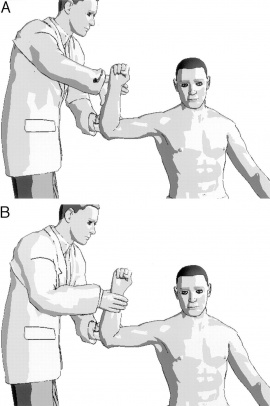
How do you do this test[11]?
You have to stand behind the patient. His injured arm is in a position of 90° abduction and 80° of external rotation. (See picture). You have to ask the patient to resist maximally the first external rotation (A) and then the internal rotation (B). Here you are testing manually the isometric muscle for the external rotation and internal rotation. If the patient has a good strength in external rotation and a weakness while doing the internal rotation, than you can say that the IRRST is positive.
A positive IRRST is based on the fact that at a fixed position of the arm, the medial rotators the caput humerus will do a translocation to the front against the frontal labrum.
A positive result is predictive for an internal impingement, that is frequently a Bankart lesion, and a negative test would suggest a classic outlet impingement[12]
Medical Management
[edit | edit source]
Arthroscopic repair with suture anchors is an effective surgical technique for the treatment of an isolated Bankart lesion. Open repair does not always show us a significantly improvement of functionality of the shoulder. [5][6][13] Identification of risk factors for recurrence allows for appropriate patient counseling and consideration of open stabilization. Risk factors are having an age under age 25, ligamentous laxity, and with a large (>250 mm³) Hill-Sachs lesion [14]. After arthroscopic surgery there are still one third of the stabilized shoulders that experienced still at least one dislocation after 8 to 10 years after surgery. This dislocation has a negative effect on the functionality of the shoulder[7]
Arthroscopic repair contains: A suture anchor is placed medially to the fracture on the glenoid neck, and its sutures are passed around the bony fragment through the soft tissue including the inferior glenohumeral ligament complex. The sutures of this anchor are loaded in a second anchor that is placed on the glenoid face. This creates a nontilting 2-point fixation that compresses the fragment into its bed. By use of the standard technique, additional suture anchors are used superiorly and inferiorly to the bony Bankart piece to repair the labrum and shift the joint capsule. [8] This way of surgery successful restores the shoulder stability with a high patient satisfaction.[9]
After a failed arthroscopic Bankart repair, open surgery shows us u positive outcome, including a low recurrence rate and reliable functional return. [2] Unless after open surgery there is a loss of the external rotation component. [4]
For a nonoperative treatment, there is no significant difference between IAL (intra-articular, lidocaine) and IVAS (intravenous analgesia with or without sedation). However, compared to IVAS, IAL may be less expensive, shorter recovery time, and may be associated with fewer adverse effects. [3]
Physical Therapy Management
[edit | edit source]
There are several options for interventions to the Bankart lesion. First of all we can make the difference between the operative and non-operative interventions.
Possible operative interventions include the arthroscopic Bankart repair and an open Bankart repair. In arthroscopic Bankart repair the muscle strength is regained faster, but the recurrence rates after open Bankart repair are significantly lower.[7] It is proven by several studies that recurrence rate after operative Bankart treatment is significantly reduced compared to a non-operative treatment. [13] After surgery there is of course rehabilitation needed which can be slightly comparable to the non-operative rehabilitation program. [14]
The conservative non-operative Bankart treatment had a significantly worse result with recurrent instability rates ranging from 17% to 96% in patients under age of 30 years. [13]
Rehabilitation
There are 7 key factors that need to be considered in the rehabilitation of the unstable shoulder. These are:
• Onset of the pathology (traumatic, chronic)
• Degree of instability
• Frequency of dislocation
• Direction of instability (anterior, posterior, multidirectional)
• Concomitant pathologies
• End range neuromuscular control
• Premorbid activity level [4]
Besides these key factors physiotherapist should also keep in mind that every patient is different and that we should never forget to personalize a rehabilitation program.
Focus of the rehabilitation program is on maximizing dynamic stability, scapula positioning, proprioception and improving neuromuscular control, as there are no specific exercises to improve the labrum quality. [4][8] Usually the rehabilitation program is divided in 3 phases. The programs for non-operative treatment and postoperative rehabilitation are very similar.
The first phase of rehabilitation consists of sling immobilization with a limited active range of motion for 0 to 4 weeks, this allows 20o of abduction and 40o of internal rotation. [15] This provides an earlier return to functional activity. [4][13][7][9] Immobilization in external rotation reduces the risk of recurrent shoulder dislocations. [14] After the 14 days passive movement is initiated in a pain free zone. Strengthening exercises are started as isometric contractions to initiate muscle recruitment of the rotator cuff muscles, mostly exercises in a closed kinetic chain, such as pushing your underarm against a wall towards exorotation. [8][13][4] The goals are to diminish pain and protect healing soft tissues.[4][9]
In the second phase a progressive passive motion is important, together with active-assisted range of motion exercises. [4][13] Strengthening of rotator cuff muscles is initiated in balanced exercises. Examples of exercises are movements of the shoulder executed with elastic bands or dumbbells as dynamic open chain strengthening exercises. [9] Rehabilitation should include both closed and open chain exercises. An example of a closed kinetic chain exercise is quadruped position with scapula protraction, progressing to tripod position. A patient can continue to phase three when a normal passive range of motion is achieved.
The third phase focuses on restoration of a full active range of motion. In this phase a progressively increasing resistance in dynamic exercises is stressed to regain full strength for ADL activities. Most imported in this phase is the return to full active activity of normal life. [13][9]
Key Research[edit | edit source]
add links and reviews of high quality evidence here (case studies should be added on new pages using the case study template)
Resources
[edit | edit source]
add appropriate resources here
Clinical Bottom Line[edit | edit source]
add text here
Recent Related Research (from Pubmed)[edit | edit source]
see tutorial on Adding PubMed Feed
Extension:RSS -- Error: Not a valid URL: Feed goes here!!
References[edit | edit source]
see adding references tutorial.
- ↑ Widjaja A, Tran A, Bailey M, Proper S (2006). Correlation between Bankart and Hill-Sachs lesions in anterior shoulder dislocation. ANZ J Surg 76 (6): 436–8.
- ↑ 2.0 2.1 2.2 2.3 2.4 2.5 2.6 Magnetic Resonance Imaging in Orthopaedics and Sports Medicine, Volume II, David W. Stoller, 2007, p 1329-1338
- ↑ 3.0 3.1 3.2 D. Y. Wen, ‘Current concepts in the treatment of anterior shoulder dislocations’, American Journal of Emergency Medicine, Volume 17, Number 4, 1999, p 401-407 (Level of Evidence 2A)
- ↑ 4.0 4.1 4.2 4.3 4.4 4.5 4.6 4.7 4.8 4.9 K. E. Wilk, L. C. Macrina, M. Reinold, ‘Non-operative Rehabilitation for traumatic and atraumatic glenohumeral instability’, North American Journal of Sports Fhysical Therapy, 2006, p 16-31 (Level of Evidence 1A)
- ↑ 5.0 5.1 5.2 S. Waldt, A. Burkart, A. B. Imhoff, M. Bruegel, E. J. Rummeny, K. Woerler, ‘Anterior Shoulder Instability: Accuracy of MR Arthrography in the Classification of Anteroinferior Labroligamentous Injuries’, Radiology, Volume 237, Number 2, 2005, p 578-583 (Level of Evidence 2B)
- ↑ 6.0 6.1 J. Beltran, Z. S. Rosenberg, V. P. Chandnani, F. Cuomo, S. Beltran, A. Rokito, ‘Glenohumeral Instability: Evaluation with MR Arthrography’, Scientific Exhibit, Volume 17, Number 3 (Level of Evidence 2C)
- ↑ 7.0 7.1 7.2 7.3 7.4 F. KHIAMI, ET AL. (February 2012), Anterior shoulder instability arthroscopic treatment outcomes measures: The WOSI correlates with the Walch-Duplay score, Revue de chirurgie Orthopédique et Traumatologique, volume 98, pages 48-53fckLRLevel of evidence: 4
- ↑ 8.0 8.1 8.2 8.3 DOMINIQUE M. ET AL (December 27), Validation of the Instability Shoulder Index Score in a Multicenter Reliability Study in 114 Consecutive cases, Am J Sports Med 2013 41: 278fckLRLevel of evidence: 2B
- ↑ 9.0 9.1 9.2 9.3 9.4 9.5 T. DUNCAN TENNENT,* FRCS(ORTH), WILLIAM R. BEACH, MD, AND JOHN F. MEYERS, MD, A Review of the Special Tests Associated with Shoulder Examination Part I: The Rotator Cuff Tests. Vol. 31, No. 1, 2003. pag 157-158.fckLRLevel of evidence: 3A
- ↑ M.H. Moen, R.-J. de vos, E.R.A. van Arkel, A. Weir, J. Moussavi, T. Kraan, D.C. de Winter: De meest waardevolle klinische schoudertesten. Sport en geneeskunde. Oktober 2008, nummer 4. P6-10
- ↑ Zaslav KR. Internal rotation resistance strength test: a new diagnostic test to differentiate intra-articular pathology from outlet (Neer) impingement syndrome in the schoulder. J. Shoulder Elbow Surg 2001 Jan-Feb;10(1):23-7.
- ↑ K. Van Nugteren, D. Winkel, P. van der Tas. Van, Onderzoek en behandeling van de schouder: orthopedisch casuistiek. Maart 2007, hoofdstuk 4, p43-47. Level Vf
- ↑ 13.0 13.1 13.2 13.3 13.4 13.5 13.6 NETTO ET AL ; Treatment of Bankart Lesions in Traumatic Anterior Instability of the Shoulder: A Randomized Controlled Trial Comparing Arthroscopy and Open Techniques; 2012fckLRLevel of evidence: 2
- ↑ 14.0 14.1 14.2 VOOS ET AL. Prospective Evaluation of Arthroscopic Bankart Repairs for Anterior Instability;. 2009 fckLRLevel of evidence: 4
- ↑ S-H Kim, K-I Ha, M-W Jung, M-S Lim, Y-M Kim, J-H Park, ‘Accelerated Rehabilitation After Arthroscopic Bankart Repair for Selected Cases: A Prospective Randomized Clinical Study’, Arthroscopy, 2003, Volume 19, Number 7 (Level of Evidence 1B)

