Avulsion Fractures of the Ankle
Original Editors - Niels Verbeeck
Top Contributors - Niels Verbeeck, Kim Jackson, Admin, Pierreux Maarten, Shaimaa Eldib, Scott Cornish, Uchechukwu Chukwuemeka, Vidya Acharya, Rachael Lowe, Jan Alderweireldt, Claire Knott, Wanda van Niekerk, Lucinda hampton and 127.0.0.1
Definition/Description
[edit | edit source]
An avulsion fracture is a fracture caused by a traumatic traction (longterm repetitive pulling or single high impact traumatic traction) from a tendon to the bone. Because the tendon can bear more pull than the bone, it is common that the bone will pull off instead of the tendon. The tendon pulls off a tiny fragment of bone.[1][2]
An avulsion fracture can occur at many places in the body, but some areas in the body are more sensitive than others. An avulsion fracture of the ankle mostly occur at the lateral of medial malleolus. In the foot the most common is at the base of the fifth metatarsal, but may also be found at the talus and calcaneus. A twisting injury to the ankle and foot may cause an avulsion fracture in any of these locations.
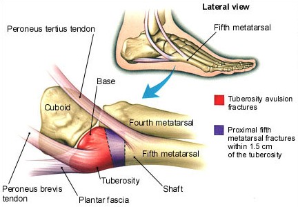
Clinically Relevant Anatomy[edit | edit source]
The 5th metatarsal is located at the lateral side of the foot. The metatarsal is divided into 3 parts: the base (also called tuberosity), the metaphysis and the head. The peroneus (fibularis) brevis’ is attached at the lateral side of the tuberosity metatarsal V.(red area at figure) The peroneus (fibularis) tertius’ is attached at the dorsal side of the most proximal compartment of the metaphysis. Because of large traction by these structures, during inversion injury, tuberosity avulsion fractures occur. (between red and purple area at figure)
figure1: source: http://www.med-info.nl/images/images_trauma/Trauma_voet_MT5_Jones_groot.jpg
Characteristics/Clinical Presentation[edit | edit source]
The clinical characteristics of avulsion fracture are different from those of ligament rupture. Unlike nonoperative treatment of lateral ligament rupture, nonoperative treatment of avulsion fracture does not yield satisfactory results. Symptoms of an ankle avulsion fracture are very similar to an ankle sprain and it is very difficult to tell the difference without an X-ray or MRI scan. There will be pain in the ankle immediately after the injury occurs with immediate swelling. Bruising may develop later and the athlete will most likely have difficulty moving or putting weight on the ankle. A patient who suffers an acute rolling of their ankle can also injure the base of the 5th metatarsal. This will produce immediate pain over the outside aspect of the foot. It can be associated with significant swelling. Over time, the skin can turn black and blue. It will be associated with quite specific local tenderness over the base of the bone on the outside of the foot (the 5th metatarsal). Patients who have suffered a 5th metatarsal base avulsion fracture will give a history of a twisting injury to their ankle and foot (inversion plantarflexion injury) similar to what occurs with an ankle sprain. [3][4][5]
Differential Diagnosis[edit | edit source]
Avulsion fractures are often confused with other types of fractures to the fifth metatarsal:
- A Jones fracture occurs as a result of a stress fracture, due to repetitive loading of the outside part of the foot from the patient’s underlying foot pattern or lower extremity alignment. Unlike a Dancer’s fracture a Jones fracture may not heal and often requires surgery.[6]
- Stress fractures
- Mid-shaft fractures
Diagnostic Procedures[edit | edit source]
An x-ray may be ordered by the surgeon. Avulsion fractures are sometimes overlooked when an x-ray is taken or when the injury to MT V occurs together with an ankle sprain. In this case other imaging studies are recommended, such as MR imaging, CT scan or scintigrams. [2] [7] [8]
Outcome Measures[edit | edit source]
Good predictors of outcome are a splint made plaster and periods of no weight bearing (NWB). The evaluation of this happens with using Olerud ankle score, with linear analogue scales (LAS) for pain and comfort, and with questions about cosmesis and wearing of shoes. Gender, age, and fracture type did not affect outcome. The pain scale ranged from 0 (no pain) to 10 (the worst pain one could imagine), and the comfort scale also ranged from 0 (the least possible comfort) to 10 (perfect comfort).[9]
add links to outcome measures here (also see Outcome Measures Database)
Examination[edit | edit source]
During the medical history taking the surgeon or a physiotherapist has to ask the patient how the injury occurred and when the pain started. Using the Ottawa Ankle Rules localize the exact area of pain. Palpation of the 5th metatarsal tuberosity is painfull.
Medical Management[edit | edit source]
Patients with an avulsion fracture of the base of the 5th metatarsal are usually treated conservative. If the bone is not displaced, the treatment is accomplished with a walking boot or a walking cast. They will be casted for four to six weeks. Surgery is only recommended if the bone is displaced from its normal position or when there is more than 30% of the cubometatarsal joint involved. The bone will be removed or fixed with osteosynthesis material. Crutches may be useful to avoid weight on the injured foot. [10]
Physical Therapy Management
[edit | edit source]
Avulsion fractures are often treated as ankle sprains. A physiotherapist doesn’t treat ‘an avulsion fracture’ on its own, but the dysfunctional movement and impairments of an individual. Therefore it is important to individualize the treatment. [8]
It is important to follow a full ankle rehabilitation after an avulsion fracture, because inappropriately managed avulsion fractures can lead to significant long-term functional disability. Most fractures heal well, but following a strict immobilization it is indicated to regain full range of motion (normal arthrokinematics), strength of the lower extremity muscles, proprioception and functionality in sport.
A full ankle rehabilitation after an avulsion fracture consists of three phases, the acute phase, the recovery phase and the functional phase:
- The first phase, also called ‘the acute phase’, can be started at two weeks postoperatively. The first phase will include passive range of motion exercises and crytherapy. This phase is based on the reduction of pain, inflammation and edema while retarding muscle atrophy of the lower extremity complex.
- The second phase, also called ‘the recovery phase’,can be started after the patient has met the goals of the first phase.
We can divide the rehabilitation program in 3 phases:
- During the first 2 weeks: we will start with active range of motion exercises for the toes and the MPT joints, strengthening exercises for the ankle and foot are still too early. By the second week, isometric exercises to the dorsiflexors, plantar flexors, invertors and evertors of the foot are started. Active ankle movements are begun.
- After 2 weeks: By week 6-8, we can start with active and passive range of motion exercises fot the ankle and the subtalar joint. Isometric and isotonic exercises for the ankle and subtalar joint are advised. Exercises improving lower extremity strength with theraband and proprioception exercises with a biomechanical ankle platform system. At the beginning of the second phase, advice the patients they should use the least resistant band. Toward the end of the second phase, the patient should begin using a wobble board to improve proprioception and begin closed kinetic chain activities (walking and loading).
- After 8-12 weeks: strengthening exercises to the dorsiflexors, plantarflexors, invertors, evertors, long flexors and extensors of the toes. Full weight-bearing exercises may be permitted.[11][12] - The third phase, also called ‘the functional phase’, can be started at six to eight weeks postoperatively. The third phase involves increasing power of the lower extremity complex, increasing neuromuscular control and utilizing sport-specific training of the lower extremity for a full return to sport. [13]
Resources
[edit | edit source]
Articles:
- E.W. Zwitser *, R.S. Breederveld, Fractures of the fifth metatarsal; diagnosis and treatment, Injury, Int. J. Care Injured 41 (2010) 555–562 (level: A1)
- Haraguchi N, Toga H, Shiba N, Kato F, Avulsion fracture of the lateral ankle ligament complex in severe inversion injury: incidence and clinical outcome, Am J Sports Med. 2007 Jul;35(7):1144-52 (level: A2)
- Peter Vorlat , Wim Achtergael , Patrick Haentjens, Predictors of outcome of non-displaced fractures of the base of the fifth metatarsal, International Orthopaedics (SICOT) (2007) 31: 5–10 (level: B)
- Duke G. Pao, Theodore E. Keats, Robert G. Dussault, Avulsion Fracture of the Base of the Fifth Metatarsal Not Seen on Conventional Radiography of the Foot: The Need for an Additional Projection, AJR 2000;175:549–552 (level: C)
Books:
- David F. Paton. Fractures and Orthopaedics. Edinburgh: Churchill Livingstone, 1988.
- Ronald McRace. Practical Fracture Treatment. 3rd ed. Edinburgh: Churchill Livingstone, 1994.
Sites:
- http://orthopedics.about.com/od/brokenbones/a/avulsion.htm
- http://www.foothealthfacts.org/footankleinfo/fifth-metatarsal_fractures.htm
- http://www.podiatrytoday.com/article/6565
- http://www.epainassist.com/sports-injuries/ankle-injuries/ankle-avulsion-fracture-symptoms-causes-treatment
References[edit | edit source]
- ↑ http://orthopedics.about.com/od/brokenbones/a/avulsion.htm
- ↑ 2.0 2.1 http://www.foothealthfacts.org/footankleinfo/fifth-metatarsal_fractures.htm
- ↑ Fracture Dislocations of the Tarsometatarsal Joints: End Results Correlated with Pathology and Treatment Level of evidence: 2A
- ↑ Haraguchi N, Toga H, Shiba N, Kato F, Avulsion fracture of the lateral ankle ligament complex in severe inversion injury: incidence and clinical outcome, Am J Sports Med. 2007 Jul;35(7):1144-52
- ↑ http://www.epainassist.com/sports-injuries/ankle-injuries/ankle-avulsion-fracture-symptoms-causes-treatment
- ↑ http://www.epainassist.com/sports-injuries/ankle-injuries/ankle-avulsion-fracture-symptoms-causes-treatment
- ↑ Duke G. Pao, Theodore E. Keats, Robert G. Dussault, Avulsion Fracture of the Base of the Fifth Metatarsal Not Seen on Conventional Radiography of the Foot: The Need for an Additional Projection, AJR 2000;175:549–552
- ↑ 8.0 8.1 Haraguchi N, Toga H, Shiba N, Kato F, Avulsion fracture of the lateral ankle ligament complex in severe inversion injury: incidence and clinical outcome, Am J Sports Med. 2007 Jul;35(7):1144-52
- ↑ http://www.footeducation.com/foot-and-ankle-conditions/dancers-fracture-5th-metatarsal-avulsion-fracture
- ↑ E.W. Zwitser *, R.S. Breederveld, Fractures of the fifth metatarsal; diagnosis and treatment, Injury, Int. J. Care Injured 41 (2010) 555–562
- ↑ Essentials of Orthopaedics for Physiotherapist, Ebnezar Level of evidence:E
- ↑ Fracture Dislocations of the Tarsometatarsal Joints: End Results Correlated with Pathology and Treatment Level of evidence: 2A
- ↑ http://www.podiatrytoday.com/article/6565

