Ankle Sprain: Difference between revisions
No edit summary |
No edit summary |
||
| Line 151: | Line 151: | ||
*Taping and making an appointment for a check-up to evaluate the healing of the ankle sprain<ref name="kngf0">Van der Wees PJ, Lenssen AF, Feijts YAEJ, Bloo H, van Moorsel SR, Ouderland R, et al. KNGF-Guideline for Physical Therapy in patients with acute ankle sprain. Dutch J Phys Ther. 2006; 116(Suppl 5):**. Available from: https://www.kngfrichtlijnen.nl/images/imagemanager/guidelines_in_english/KNGF_Guideline_for_Physical_Therapy_in_patients_with_Acute_Ankle_Sprain.pdf (accessed 29 Aug 2012). </ref><ref name="Allen"> Fongemie A, Dudero A, Standemo G, Stovitz S, Dahm D, THomas A, et al. Health Care Guideline [Internet]. Institute for Clinical Systems Improvement. Health care guideline: ankle sprain. 7th ed. 2006. Available from: http://www.icsi.org/ankle_sprain/ankle_sprain_4.html (accessed 29 Aug 2012)</ref> | *Taping and making an appointment for a check-up to evaluate the healing of the ankle sprain<ref name="kngf0">Van der Wees PJ, Lenssen AF, Feijts YAEJ, Bloo H, van Moorsel SR, Ouderland R, et al. KNGF-Guideline for Physical Therapy in patients with acute ankle sprain. Dutch J Phys Ther. 2006; 116(Suppl 5):**. Available from: https://www.kngfrichtlijnen.nl/images/imagemanager/guidelines_in_english/KNGF_Guideline_for_Physical_Therapy_in_patients_with_Acute_Ankle_Sprain.pdf (accessed 29 Aug 2012). </ref><ref name="Allen"> Fongemie A, Dudero A, Standemo G, Stovitz S, Dahm D, THomas A, et al. Health Care Guideline [Internet]. Institute for Clinical Systems Improvement. Health care guideline: ankle sprain. 7th ed. 2006. Available from: http://www.icsi.org/ankle_sprain/ankle_sprain_4.html (accessed 29 Aug 2012)</ref> | ||
First time lateral ligament sprains can be innocuous injuries that resolve quickly with minimal intervention and some approaches suggest that minimal intervention can be given to this population. The NICE guidelines 2016 recommend advice and analgesia, but not routine physiotherapy referrals<ref>NICE, 2016. [https://cks.nice.org.uk/sprains-and-strains#!scenario Sprains and Strains]. https://cks.nice.org.uk/sprains-and-strains#!scenario [accessed 5 January 2016]</ref>. However it has has also been highlighted that recurrence rate of first time lateral ankle sprains is 70%<ref>Sefton JM, Hicks-Little CA, Hubbard TJ, Clemens MG, Yengo CM, Koceja DM, Cordova ML. Sensorimotor function as a predictor of chronic ankle instability. Clinical Biomechanics. 2009 Jun 30;24(5):451-8.</ref>. With the recurrence rate so high and the guidelines not recommending any rehabilitation others have questioned this approach<ref name="Docherty 2">Doherty C, Bleakley C, Hertel J, Caulfield B, Ryan J, Delahunt E. [http://journals.sagepub.com/doi/abs/10.1177/0363546516628870?url_ver=Z39.88-2003&amp;amp;amp;amp;amp;rfr_id=ori%3Arid%3Acrossref.org&amp;amp;amp;amp;amp;rfr_dat=cr_pub%3Dpubmed&amp;amp;amp;amp;amp; Recovery From a First-Time Lateral Ankle Sprain and the Predictors of Chronic Ankle Instability A Prospective Cohort Analysis. The American journal of sports medicine]. 2016 Apr 1;44(4):995-1003</ref>. | First time lateral ligament sprains can be innocuous injuries that resolve quickly with minimal intervention and some approaches suggest that minimal intervention can be given to this population. The NICE guidelines 2016 recommend advice and analgesia, but not routine physiotherapy referrals<ref>NICE, 2016. [https://cks.nice.org.uk/sprains-and-strains#!scenario Sprains and Strains]. https://cks.nice.org.uk/sprains-and-strains#!scenario [accessed 5 January 2016]</ref>. However it has has also been highlighted that recurrence rate of first time lateral ankle sprains is 70%<ref>Sefton JM, Hicks-Little CA, Hubbard TJ, Clemens MG, Yengo CM, Koceja DM, Cordova ML. Sensorimotor function as a predictor of chronic ankle instability. Clinical Biomechanics. 2009 Jun 30;24(5):451-8.</ref>. With the recurrence rate so high and the guidelines not recommending any rehabilitation others have questioned this approach<ref name="Docherty 2">Doherty C, Bleakley C, Hertel J, Caulfield B, Ryan J, Delahunt E. [http://journals.sagepub.com/doi/abs/10.1177/0363546516628870?url_ver=Z39.88-2003&amp;amp;amp;amp;amp;amp;rfr_id=ori%3Arid%3Acrossref.org&amp;amp;amp;amp;amp;amp;rfr_dat=cr_pub%3Dpubmed&amp;amp;amp;amp;amp;amp; Recovery From a First-Time Lateral Ankle Sprain and the Predictors of Chronic Ankle Instability A Prospective Cohort Analysis. The American journal of sports medicine]. 2016 Apr 1;44(4):995-1003</ref>. | ||
=== Severe Ankle Sprain === | === Severe Ankle Sprain === | ||
| Line 270: | Line 270: | ||
|- | |- | ||
| {{#ev:youtube|PDFbZFNtPfs|260}} <ref>Coordinated Health TV. Ankle Sprains Part 1: Anatomy. Available from: https://www.youtube.com/watch?v=PDFbZFNtPfs[last accessed 24/03/2015]</ref> | | {{#ev:youtube|PDFbZFNtPfs|260}} <ref>Coordinated Health TV. Ankle Sprains Part 1: Anatomy. Available from: https://www.youtube.com/watch?v=PDFbZFNtPfs[last accessed 24/03/2015]</ref> | ||
| {{#ev:youtube|dP17ZY3zxa4|260}} <ref>Coordinated Health TV. Ankle Sprains Part 2: Symptoms &amp;amp;amp;amp;amp;amp;amp;amp;amp;amp;amp;amp;amp;amp;amp;amp;amp;amp;amp;amp;amp;amp;amp;amp;amp;amp;amp;amp;amp;amp;amp;amp;amp;amp;amp;amp; Evaluation. Available from: https://www.youtube.com/watch?v=dP17ZY3zxa4 [last accessed 24/03/2015]</ref> | | {{#ev:youtube|dP17ZY3zxa4|260}} <ref>Coordinated Health TV. Ankle Sprains Part 2: Symptoms &amp;amp;amp;amp;amp;amp;amp;amp;amp;amp;amp;amp;amp;amp;amp;amp;amp;amp;amp;amp;amp;amp;amp;amp;amp;amp;amp;amp;amp;amp;amp;amp;amp;amp;amp;amp;amp; Evaluation. Available from: https://www.youtube.com/watch?v=dP17ZY3zxa4 [last accessed 24/03/2015]</ref> | ||
| {{#ev:youtube|dznWBbwLq6k|260}} <ref>Coordinated Health TV. Ankle Sprains Part 3: Rehab &amp;amp;amp;amp;amp;amp;amp;amp;amp;amp;amp;amp;amp;amp;amp;amp;amp;amp;amp;amp;amp;amp;amp;amp;amp;amp;amp;amp;amp;amp;amp;amp;amp;amp;amp;amp; Protection. Available from: https://www.youtube.com/watch?v=dznWBbwLq6k[last accessed 24/03/2015]</ref> | | {{#ev:youtube|dznWBbwLq6k|260}} <ref>Coordinated Health TV. Ankle Sprains Part 3: Rehab &amp;amp;amp;amp;amp;amp;amp;amp;amp;amp;amp;amp;amp;amp;amp;amp;amp;amp;amp;amp;amp;amp;amp;amp;amp;amp;amp;amp;amp;amp;amp;amp;amp;amp;amp;amp;amp; Protection. Available from: https://www.youtube.com/watch?v=dznWBbwLq6k[last accessed 24/03/2015]</ref> | ||
|} | |} | ||
| Line 356: | Line 356: | ||
<br> | <br> | ||
[[Category:Interventions | [[Category:Interventions]][[Category:Sports_Injuries]][[Category:Musculoskeletal/Orthopaedics|Orthopaedics]][[Category:Vrije_Universiteit_Brussel_Project]][[Category:Foot_and_Ankle_Conditions]][[Category:Ankle]] | ||
Revision as of 12:45, 29 March 2017
Original Editor - Dale Boren, Michael Kauffmann, Pieter Jacobs
Top Contributors - Naomi O'Reilly, Laura Ritchie, Admin, Rachael Lowe, Michael Kauffmann, Kim Jackson, Pieter Jacobs, Reem Ramadan, Cath Young, Alex Palmer, Roberto Monfermoso, Dale Boren, Andrew Costin, Adam Vallely Farrell, Corentin Meese, 127.0.0.1, Scott Cornish, Rucha Gadgil, Scott Buxton, Anas Mohamed, Khloud Shreif, Nupur Smit Shah, WikiSysop, Asha Bajaj, Simisola Ajeyalemi, Lisa Parijs, Candace Goh, Evan Thomas, Shaimaa Eldib, Wanda van Niekerk, Anouck Leo, Olajumoke Ogunleye, Kai A. Sigel, Fasuba Ayobami and Tony Lowe
Definition/Description[edit | edit source]
An ankle sprain is where one or more of the ligaments of the ankle are partially or completely torn.
Epideimiology[edit | edit source]
An ankle sprain is a common injury. Inversion ankle sprains are the most common, making up 85% of all ankle sprains. It is know that the incidence of lateral ligament injuries is the most common amongst the sporting population and the consequence of not rehabilitating after an initial injury increases the chances of recurrence[1].
Clinically Relevant Anatomy[edit | edit source]
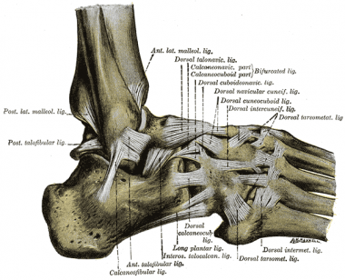
The most commonly torn ankle ligament is the anterior talofibular ligament (ATFL) which is on the lateral aspect of the ankle. Additional ligaments on the lateral aspect of the ankle include the calcaneo-fibular ligament (CFL) and the posterior talofibular ligament (PTFL).
| [2] |
On the medial side of the ankle the strong deltoid ligament complex which consists of the posterior tibiotalar ligament, tibiocalcaneal ligament, tibionavicular ligament and anterior tibiotalar ligament may be injured with forceful eversion.
A syndesmotic (high ankle) sprain may occur with external rotation with dorsiflexion. It involves the Distal Tibiofibular Syndesmosis and may injure the anterior-inferior tibiofibular ligament, posterior-inferior tibiofibular ligament, transverse tibiofibular ligament, interosseous membrane, interosseous ligament, inferior transverse ligament.
Mechanism of Injury / Pathological Process[edit | edit source]
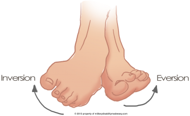
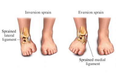
The most common mechanism of injury for an ankle sprain is an inversion injury commonly called a lateral or inversion ankle sprain. This is when the foot is forced into a combined movement of plantarflexion and inversion, which applies too much force to the lateral structures of the ankle; causing them to fail[1]. A less common mechanism of injury involves a forceful eversion movement at the ankle, resulting in injury to the strong deltoid ligament.
| Aspect | Mechanism of injury | Ligaments |
|---|---|---|
| Lateral | Inversion and plantarflexion | anterior talofibular ligament calcaneo-fibular ligament posterior talofibular ligament |
| Medial | Eversion | posterior tibiotalar ligament tibiocalcaneal ligament tibionavicular ligament anterior tibiotalar ligament |
| High | External rotation and dorsiflextion | anterior-inferior tibiofibular ligament posterior-inferior tibiofibular ligamen transverse tibiofibular ligament interosseous membrane interosseous ligament inferior transverse ligament |
Clinical Presentation[edit | edit source]
- Presents with history of inversion injury or forceful eversion injury to the ankle. May have previous history of ankle injuries or instability.
- Able to weight-bear through the limb.
- If patient presents with description of cold foot or paraesthesia, suspect neurovascular compromise of peroneal nerve.
- Tenderness, swelling and bruising can occur on either side of the ankle.
- No bony tenderness, deformity or crepitus present.
- Passive inversion or plantar flexion + inversion should replicate symptoms for a lateral ligamnet sprain, passive eversion should replicate symproms for a medial ligamnet sprain.
- Special Tests: +ve Anterior Draw, Talar Tilt or Squeeze Test (depending structures involved).
Differential Diagnosis
[edit | edit source]
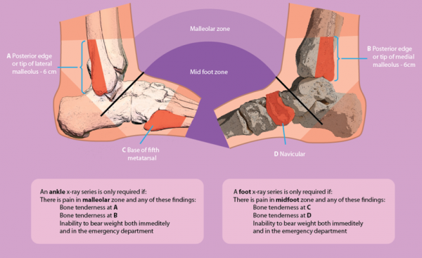
The Ottawa Ankle Clinical Prediction Rules constitute an accurate tool to exclude fractures within the first week after an ankle injury.[3]
Additional differential diagnosis to look out for:[4]
- Impingement
- Tarsal Tunnel Syndrome
- Sinus Tarsi Syndrome
- Cartilage or Osteochondral Injuries
- Peroneal Tendinopathy or subluxation
- Posterior Tibial Tendon Dysfunction
Classification Grading Systems[edit | edit source]
There are a number of different grading systems used for the classification of ligament sprains each have strengths and weaknesses. One important consideration is that each therapists will employ different systems so it is important to be aware of a wide variety for continuity of care. This is evident when reading research regarding sprains and authors not disclosing which system they used, reducing rigour and quality of the write up of research[5].
The traditional grading system for ligament injuries focuses on a single ligament[5]
- Grade I injury represents a microscopic injury without stretching of the ligament on a macroscopic level.
- Grade II injury has macroscopic stretching, but the ligament remains intact.
- Grade III injury is a complete rupture of the ligament.
As all ready discussed in ligaments of the ankle the ankle has several complexes of multiple ligaments so it may be difficult and inappropriate to use a system that is suited for the description of the state of a single ligament unless it can be sure only one ligament it torn.
Some authors have thus resorted to grading lateral ankle-ligament sprains by the number of ligaments injured[6]. Again it is hard to be certain on the number of ligaments torn unless there is clear, high quality radiographic or surgical evidence.
A third system which can be adopted is a 3 graded classification based on the severity of sprain injury[5].
- Grade I - Mild - Little swelling & tenderness with little impact on function
- Grade II - Moderate - Moderate swelling, pain and impact on function. Reduced proprioception, ROM and instability
- Grade III - Severe - Complete rupture, large swelling, tenderness+++, Loss of function and marked instability
This scale is largely subjective due to individual therapist interpretation. However the same can be said for the other classifications unless clear radiographic evidence is available or assessment and treatment by surgical intervention. In this case the assessment will be post-treatment.
Outcome Measures[edit | edit source]
Lower Extremity Functional Scale (LEFS)
For other outcome measures see: Outcome Measures Database
Clinical Examination
[edit | edit source]
In the case of an ankle sprain multiple structures may be involved therefore a full foot and ankle assessment is recommended[5].
The therapist should begin by taking a thorough subjective history to gain an understanding of the mechanism of injury. The therapist should observe gait pattern, standing posture and the position of wear on the individuals shoes.[7] The assessor should observe for any gross deformity, malalignment or atrophy of the musculature. Any oedema and ecchymosis should be noted.
The foot, ankle and lower leg should be palpated to feel the structures that may be involved in the injury (including bone, muscle and ligamentous structures). Following which, active and passive range of movement should be assessed.
| [8] | [9] |
Special Tests[edit | edit source]
- Anterior Draw - tests the ATFL
- Talar Tilt - tests the CFL
- Posterior Draw - tests the PTFL
- Squeeze test - for syndesmotic sprain
- External rotation stress test (Kleiger’s test) - syndesmotic sprain
| [10] | [11] |
It has been suggested that these tests be performed at 4-7 days post acute injury to allow the initial swelling and pain to settle, this enable the clinician to gain a more accurate diagnosis[5].
Physical Therapy Management[edit | edit source]
Mild Ankle Sprain[edit | edit source]
- Natural full recovery within 14 days
- Taping and making an appointment for a check-up to evaluate the healing of the ankle sprain[3][12]
First time lateral ligament sprains can be innocuous injuries that resolve quickly with minimal intervention and some approaches suggest that minimal intervention can be given to this population. The NICE guidelines 2016 recommend advice and analgesia, but not routine physiotherapy referrals[13]. However it has has also been highlighted that recurrence rate of first time lateral ankle sprains is 70%[14]. With the recurrence rate so high and the guidelines not recommending any rehabilitation others have questioned this approach[15].
Severe Ankle Sprain[edit | edit source]
Physiotherapy is required. Functional therapy of the ankle is shown to be more efficient than immobilisation. Functional therapy treatment will be divided in four steps, related to the four steps in the tissue recovery after an acute ankle sprain, [3] Inflammatory phase, Proliferative phase, Early Remodelling, Late Maturation and Remodelling. [3][12][16][17][18]
Step 1: Inflammatory Phase (0-3 days)[edit | edit source]
Goals:[edit | edit source]
Reduction of pain and swelling and improve circulation and partial foot support
Actions:[edit | edit source]
The most common approach to manage ankle sprain consists of Protection, Rest, Ice, Compression, and Elevation (PRICE)
1. Recommendations for the Patient:
- PROTECTION: Protect the ankle from further injury by resting and avoiding activities that may cause further injury and/or pain
- REST: Advise rest for the first 24 hours after injury, possibly with crutches to offload the injured ankle and altering work and sport and exercise requirements as needed
- ICE: Apply a cold application (15 to 20 minutes, one to three times per day)
- COMPRESSION: Apply compression bandage to control swelling caused by the ankle sprain
- ELEVATION: Ideally elevate ankle above the level of the heart but minimally avoid positions where the ankle is in a dependent position relative to the body
2. Practice Foot and Ankle Functions:
- Ask your patient to move toes and ankle within pain-free limits to improve local circulation. [12][19][20]
Step 2: Proliferative Phase (4-10 days)[edit | edit source]
Goals:[edit | edit source]
Recovery of foot and ankle function and improved load-carrying capacity.
Actions:
[edit | edit source]
1. Patient education regarding gradual increase in activity level, guided by symptoms.
2. Practise Foot and Ankle Functions
- Range of Motion
- Active Stability
- Motor Coordination
3. Tape/Brace :
- Apply tape as soon as the swelling is decreased.
- Whether you use a tape or a brace depends on the preferences of the patient.
- Boyce et al (2005) found that the use of an Aircast ankle brace for the treatment of lateral ligament ankle sprains produces a significant improvement in ankle joint function compared with standard management with an elastic support bandage.[21]
- Still it remains uncertain which treatment (brace, bandage or tape) is most beneficial. [3]
Here are two examples of ankle sprain taping techniques but please note that many different techniques exist.
| [22] | [23] |
Step 3: Early Remodelling (11 -21 days)[edit | edit source]
Goals: [edit | edit source]
Improve muscle strength, active (functional) stability, foot/ankle motion, mobility (walking, walking stairs, running).
Actions:[edit | edit source]
1. Education:
- Provide information about possible preventive measures (tape or brace)
- Advise regarding appropriate shoes to wear during sport activities - judge the quality of the shoes in relation to the type of sport and surface
2. Practise Foot and Ankle Functions (See Resource Videos below)
- Practice balance, muscle strength, ankle/foot motion and mobility (walking, stairs, running).
- Look for a symmetric walk pattern.
- Work on dynamic stability as soon as load-bearing capacity allows, focusing on balance and coordination exercises. Gradually progress the loading, from static to dynamic exercises, from partially loaded to fully loaded exercises and from simple to functional multi-tasking exercises. Alternate cycled with non-cycled exercises (abrupt, irregular exercises). Use different types of surfaces to increase the level of difficulty.
- Encourage the patient to continue practicing the functional activities at home with precise instructions regarding the expectations for each exercise.
3. Bandage
- Advise to wear tape or brace during physical activities. These procedures are needed until the patient is able to execute correctly the static and dynamic exercises of balance and motor coordination.
Step 4: Late Remodelling and Maturation[edit | edit source]
Goals:[edit | edit source]
Improve the regional load-carrying capacity, walking skills and improve the skills needed during activities of daily living as well as work and sports.
Actions:[edit | edit source]
1. Practise and Adjust Foot Abilities (Functions and Activities)
- Practise motor coordination skills while practising mobility exercises
- Continue to progress the load-bearing capacity as described above until the pre-injury load-carrying capacity is reached
- Increase the complexity of motor coordination exercises in varied situations until the pre-injury level is reached
- Encourage the patient to continue practicing at home
Chronic Ankle Instability[edit | edit source]
On going issues following a lateral ligament injury within the ankle are reported in 19-72% of patients. An ability to complete certain movement tasks, evidence of deficits during the Star Excursion Balance Test and poorer self-reported function as quantified using the Foot and Ankle Ability Measure can be utilized as predictive measures of a Chronic Ankle Instability (CAI) outcome in the clinical setting for patients with a first-time lateral ankle sprain injury[24]. Around 20% of people develop CAI and this has been attributed to a delayed muscle reflex of stabilizing lower leg muscles, deficits in lower leg muscle strength, deficits in kinaesthesia, or an impaired postural control[25][26].
It is recommended that all patients undergo conservative treatment to improve stability and improve the muscle reflex and strength of those stabilising lower limb muscles. Although this may help some people cannot compensate for defecit of the lateral ligament complex, and therefore surgery is required[25].
Read more about Chronic Ankle Instability here
Resources
[edit | edit source]
- The Sprained Ankle from Connecticut Centre for Orthopedic Surgery contains a range of resources on Ankle Sprains including patient resources and surgical techniques.
Coordinated Health TV Ankle Sprain Video Series
| [27] | [28] | [29] |
Denver-Vail Orthopedics, P.C Ankle Sprain Video Series
| [30] | [31] |
| [32] | [33] |
Recent Related Research (from Pubmed)[edit | edit source]
Failed to load RSS feed from http://www.ncbi.nlm.nih.gov/entrez/eutils/erss.cgi?rss_guid=1Zs2HUIS--l6SucJTzJWLjyN5ITBGb6BYpQLkEkg0_gv8VugVF|charset=UTF-8|short|max=10: Error parsing XML for RSS
Presentations[edit | edit source]
 |
Physical Therapy Management of Acute Ankle Sprain
This presentation was created by Rich Westrick and Cody McAndrew as part of the Regis University OMPT Fellowship. Physical Therapy Management of Acute Ankle Sprain/ View the presentation |
 |
PT in Injury Prevention: Ankle Sprains
This presentation was created by Sumesh Thomas as part of the Regis University OMPT Fellowship. PT in Injury Prevention: Ankle Sprains/ View the presentation |
 |
Current Best Evidence: Management of Chronic Ankle Sprain
This presentation was created by Cheryl Sparks as part of the Regis University OMPT Fellowship. Current Best Evidence: Management of Chronic Ankle Sprain/ View the presentation |
Error: Image is invalid or non-existent. |
Current Best Evidence: Management of Chronic Ankle Sprain
This presentation was created by Chris Kramer as part of the Regis University OMPT Fellowship. Current Best Evidence: Management of Chronic Ankle Sprain/ View the presentation |
References[edit | edit source]
- ↑ 1.0 1.1 Roos KG, Kerr ZY, Mauntel TC, Djoko A, Dompier TP, Wickstrom EA. The epidemiology of lateral ligament complex ankle sprains in National Collegiate Athletic Association sports. American journal of sports medicine. 2016.
- ↑ Dr Glass DPM. Ankle Sprain Injury Explained. Available from: http://www.youtube.com/watch?v=_u5w856Yjvg [last accessed 28/08/12]
- ↑ 3.0 3.1 3.2 3.3 3.4 Van der Wees PJ, Lenssen AF, Feijts YAEJ, Bloo H, van Moorsel SR, Ouderland R, et al. KNGF-Guideline for Physical Therapy in patients with acute ankle sprain. Dutch J Phys Ther. 2006; 116(Suppl 5):**. Available from: https://www.kngfrichtlijnen.nl/images/imagemanager/guidelines_in_english/KNGF_Guideline_for_Physical_Therapy_in_patients_with_Acute_Ankle_Sprain.pdf (accessed 29 Aug 2012).
- ↑ GP Online (2007). Differential diagnosis of common ankle injuries, Available at: http://www.gponline.com/differential-diagnosis-common-ankle-injuries/article/766219 (Accessed: 24th Aug 2014).
- ↑ 5.0 5.1 5.2 5.3 5.4 Lynch S. Assessment of the Injured Ankle in the Athlete. J Athl Train 2002 37(4) 406-412 Cite error: Invalid
<ref>tag; name "Lynch" defined multiple times with different content - ↑ Gaebler C, Kukla C, Breitenseher M J, et al. Diagnosis of lateral ankle ligament injuries: comparison between talar tilt, MRI and operative findings in 112 athletes. Acta Orthop Scand. 1997;68:286–290
- ↑ Anthony L. Ankle Physical Examination, Available at: http://orthosurg.ucsf.edu/oti/patient-care/divisions/sports-medicine/conditions/physical-examination-info/ankle-physical-examination/ (Accessed: 24 Aug 2014).
- ↑ Via Christi. Musculoskeletal Physical Exam: Ankle. Available from: https://www.youtube.com/watch?v=QiSm8rz2cmo [last accessed 24/03/2015]
- ↑ Massage Therapy Practise. Ankle Palpation. Available from: https://www.youtube.com/watch?v=uI8Z0obhpew [last accessed 24/03/2015]
- ↑ Physiotutors. Anterior Drawer Test (Ankle)⎟Lateral Ankle Sprain. Available from: https://www.youtube.com/watch?v=mZIqricuKjE
- ↑ Physiotutors. The Talar Tilt Test | Lateral Ankle Sprain. Available from: https://www.youtube.com/watch?v=UHNbm6Z3XK4
- ↑ 12.0 12.1 12.2 Fongemie A, Dudero A, Standemo G, Stovitz S, Dahm D, THomas A, et al. Health Care Guideline [Internet]. Institute for Clinical Systems Improvement. Health care guideline: ankle sprain. 7th ed. 2006. Available from: http://www.icsi.org/ankle_sprain/ankle_sprain_4.html (accessed 29 Aug 2012)
- ↑ NICE, 2016. Sprains and Strains. https://cks.nice.org.uk/sprains-and-strains#!scenario [accessed 5 January 2016]
- ↑ Sefton JM, Hicks-Little CA, Hubbard TJ, Clemens MG, Yengo CM, Koceja DM, Cordova ML. Sensorimotor function as a predictor of chronic ankle instability. Clinical Biomechanics. 2009 Jun 30;24(5):451-8.
- ↑ Doherty C, Bleakley C, Hertel J, Caulfield B, Ryan J, Delahunt E. Recovery From a First-Time Lateral Ankle Sprain and the Predictors of Chronic Ankle Instability A Prospective Cohort Analysis. The American journal of sports medicine. 2016 Apr 1;44(4):995-1003
- ↑ Van der Wees PJ, Lenssen AF, Hendriks EJM, Stomp DJ, Dekker J, de Brie RA. Effectiveness of exercise therapy and manual mobilisation in acute ankle sprain and functional instability: a systematic review. Aust J Physiother. 2006; 52:27-37. Available from: http://svc019.wic048p.server-web.com/ajp/vol_52/1/AustJPhysiotherv52i1van_der_Wees.pdf (accessed 29 Aug 2012)
- ↑ Bleakley CM, O’Connor SR, Tully MA, Rocke LG, MacAuley DC, Bradbury I et al. Effect of accelerated rehabilitation on function after ankle sprain: randomised controlled trial. BMJ. 2010; 340:c1964. Available from: http://www.bmj.com/content/340/bmj.c1964.pdf%2Bhtml(accessed 29 Aug 2012)
- ↑ Kerkhoffs GM, Rowe BH, Assendelft WJ, Kelly KD, Struijs PA, van Dijk CN. Immobilisation for acute ankle sprain. A systematic review.. Arch Orthop Trauma Surg. 2001;121(8):462-71. Available from: http://www.springerlink.com/content/knrf19kk4tvc2668/
- ↑ Bleakley CM, O'Connor S, Tully MA, Rocke LG, MacAuley DC, McDonough S. The PRICE study (Protection Rest Ice Compression Elevation): design of a randomised controlled trial comparing standard versus cryokinetic ice applications in the management of acute ankle sprain. BMC Musculoskelet Disord. 2007; 8:125. Available from: http://www.biomedcentral.com/content/pdf/1471-2474-8-125.pdf (accessed 29 Aug 2012)
- ↑ Bleakley C, McDonough S, MacAuley D. The use of ice in the treatment of acute soft-tissue injury. A systematic review of randomized controlled trials. Am J Sports Med. 2004;32(1):251-61. Available from: http://www.smawa.asn.au/_uploads/res/120_3630.pdf (accessed 29 Aug 2012)
- ↑ Boyce SH, Quigley MA, Campbell S. Management of ankle sprains: a randomised controlled trial of the treatment of inversion injuries using an elastic support bandage or an Aircast ankle brace. Br J Sports Med. 2005;39(2):91-6. Available from: http://bjsm.bmj.com/content/39/2/91.full.pdf+html (accessed 29 Aug 2012)
- ↑ Finest Physio. Finest Physio: Ankle Taping. Available from: http://www.youtube.com/watch?v=d_XlzZMSV8E [last accessed 09/12/12]
- ↑ itherapies. Mulligan Taping Techniques: Inversion Ankle Sprain. Available from: http://www.youtube.com/watch?v=TEjKhf-qDJU [last accessed 09/12/12]
- ↑ Doherty C, Bleakley C, Hertel J, Caulfield B, Ryan J, Delahunt E. Recovery from a first-time lateral ankle sprain and the predictors of chronic ankle instability: a prospective cohort analysis. The American journal of sports medicine. 2016 Apr;44(4):995-1003.
- ↑ 25.0 25.1 Al-Mohrej OA, Al-Kenani NS. Chronic ankle instability: Current perspectives. Avicenna journal of medicine. 2016 Oct;6(4):103.
- ↑ Eechaute C, Vaes P, Van Aerschot L, Asman S, Duquet W. The clinimetric qualities of patient-assessed instruments for measuring chronic ankle instability: a systematic review. BMC musculoskeletal disorders. 2007 Jan 18;8(1):1.
- ↑ Coordinated Health TV. Ankle Sprains Part 1: Anatomy. Available from: https://www.youtube.com/watch?v=PDFbZFNtPfs[last accessed 24/03/2015]
- ↑ Coordinated Health TV. Ankle Sprains Part 2: Symptoms &amp;amp;amp;amp;amp;amp;amp;amp;amp;amp;amp;amp;amp;amp;amp;amp;amp;amp;amp;amp;amp;amp;amp;amp;amp;amp;amp;amp;amp;amp;amp;amp;amp;amp;amp;amp; Evaluation. Available from: https://www.youtube.com/watch?v=dP17ZY3zxa4 [last accessed 24/03/2015]
- ↑ Coordinated Health TV. Ankle Sprains Part 3: Rehab &amp;amp;amp;amp;amp;amp;amp;amp;amp;amp;amp;amp;amp;amp;amp;amp;amp;amp;amp;amp;amp;amp;amp;amp;amp;amp;amp;amp;amp;amp;amp;amp;amp;amp;amp;amp; Protection. Available from: https://www.youtube.com/watch?v=dznWBbwLq6k[last accessed 24/03/2015]
- ↑ Denver-Vail Orthopedics. Ankle Sprains Part 1 How they occur, what ligaments are injured and initial treatment. Available from: https://www.youtube.com/watch?v=B0-n-ndTAX0[last accessed 24/03/2015]
- ↑ Denver-Vail Orthopedics. Ankle Sprains Part 2 Stretching and Range of Motion Exercises. Available from: https://www.youtube.com/watch?v=YHJbvf4TW2Y[last accessed 24/03/2015]
- ↑ Denver-Vail Orthopedics. Ankle Sprains Part 3 Stretching and Range of Motion Exercises. Available from: https://www.youtube.com/watch?v=u6xRWb9dFbU[last accessed 24/03/2015]
- ↑ Denver-Vail Orthopedics. Ankle Sprains Part 4 Proprioception - Balance. Available from: https://www.youtube.com/watch?v=AsEV5OYghSQ[last accessed 24/03/2015]
