Physiotherapy in Wilson's Disease
Introduction[edit | edit source]
Wilson's disease is a rare autosomal recessive disorder which causes excess copper build up in the body[1]. It mainly affects the brain, liver and cornea. It's incidence is 1 in 30,000 individuals, from ages 4 to 40 years[2].
Symptoms in this disease are varied and depend on the degree of involvement of organs damaged due to copper accumulation. Earlier signs and symptoms seen are hepatic (involving liver) in ~40% of patients, neurological (involving brain) in ~40%–50% and psychiatric in ~10% of patients[3]. The symptoms of interest from physiotherapy treatment point of view are the neurological and to some extent psychological. The neurological manifestations chiefly include tremor, dystonia, parkinsonism that can be seen clinically along with dysarthria, gait and posture disturbances, drooling and dysphagia[4].
Physiotherapy management[edit | edit source]
The symptoms of this disease, especially neurological, affect the activities of daily living of the patient to a great extent. Physiotherapy is of value in improving the quality of life of an individual affected with Wilson's Disease.
The cornerstone of management of Wilson's disease is copper-chelating therapy with penicillamine and trientine. It requires a time period of six months to take effect[2]. Until the medication starts working physiotherapy can help manage neurological symptoms of ataxia, dystonia and tremors. It also plays a role in prevention of contractures due to dystonia[2].
Physiotherapy interventions can be explained further by discussing recent case reports on Wilson's disease involving rehabilitation,
Case 1: Physical Therapy in Wilson's Disease : Case Report (2016)[5][edit | edit source]
This case report is about a 32yr. old male diagnosed with Wilson's disease 16 years ago.
Assessment[edit | edit source]
He was initially assessed using some specific tests as an outcome measures for understanding the effect of physiotherapy treatment protocol. The tests are as follows :
| Test | Parameter Assessed | |
|---|---|---|
| 1. | Timed Up and Go (TUG) | Fall Risk |
| 2. | 10 Meter Walk Test (10MWT) | Gait Speed |
| 3. | Clinical Test for Sensory Interaction on Balance
(mCTSIB) |
Static Balance |
| 4. | Functional Gait Assessment (FGA) | Dynamic Balance |
| 5. | Activities-specific Balance and Confidence Scale
(ABC Scale) |
Confidence in activities requiring balance |
Exercise Intervention[edit | edit source]
Frequency :
- Supervised physiotherapy was given 2 times per week for 2 months and 20 days.
- Home exercise was also continued 3 times per week.
The exercise protocol can be explained as follows :
- To improve Static Balance,
| Initial starting Exercise | Exercise Progression | |
|---|---|---|
| Type | Exercises in standing position | |
| Intensity | Standing exercises done on -
|
1. Standing Position -
2. Somatosensory weighting -
|
| Time | 4 series of 1 min. duration exercises with 3 min. breaks | |
2. To improve Dynamic Balance,
| Initial starting Exercise | Exercise Progression | |
|---|---|---|
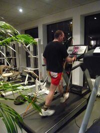
| Type | Walking on treadmill | |
| Intensity |
|
|
| Time | 3 min. with break of 5 min. ; 1 hr. per session | |
3.To improve Functional Capacity,
| Initial starting Exercise | Exercise Progression | |
|---|---|---|
| Type | Focus on daily activities which were difficult
to perform | |
| Intensity | Start with simpler exercises | Dosage according to patient's tolerance |
| Time | Rest period of 3-5 min. ; Total time 30 min. | |
Conclusion[edit | edit source]
There was improvement in patient's confidence regarding balance during various activities. The assessment tests were repeated again after the physiotherapy protocol &some showed considerable difference in pre & post values. They are as follows :
| Test | Patient Improvement
( measured in difference of pre & post test values) | |
|---|---|---|
| 1. | TUG | 3.5 seconds |
| 2. | 10 MWT
|
|
| 3. | FGA | 7 Points |
| 4. | ABC Scale | 11.2 % |
Case 2: Neuromuscular Electrical Stimulation therapy for dysphagia caused by Wilson's Disease(2012)[6][edit | edit source]
This is a case report of a 33 yrs. old male with difficulty in swallowing since last 7 years. His dysphagia has worsened over last 2-3 months. He has been diagnosed with Wilson's disease 13 years ago.
Assessment[edit | edit source]
| Test | Parameter Assessed | |
|---|---|---|
| 1. | Medical Research Council (MRC) | Limb muscle strength testing |
| 2. | Video fluoroscopic swallowing study(VFSS) | Evaluation of dysphagia according to
the functional dysphagia scale |
Intervention[edit | edit source]
| Type | Frequency | Intensity | Time | |
|---|---|---|---|---|
| 1. | Neuromuscular Electrical Nerve stimulation
(NMES) Positioning of Electric pads :
|
5 times a week for 2 weeks
Total 10 sessions |
Current Intensity dosage:
|
1 hr. per day |
Conclusion[edit | edit source]
The patient had oral & pharyngeal phase abnormalities in swallowing as assessed during VFSS pre-treatment. Post 10 sessions improvement was observed in the pharyngeal phase of swallowing with marked reduction (<10%) in vallecular residue as compared to pre-NMES VFSS.
References[edit | edit source]
- ↑ Bull PC, Thomas GR, Rommens JM, Forbes JR, Cox DW. The Wilson disease gene is a putative copper transporting P–type ATPase similar to the Menkes gene. Nature genetics. 1993 Dec;5(4):327-37.
- ↑ 2.0 2.1 2.2 Chaudhry HS, Anilkumar AC. Wilson Disease. [Updated 2021 Aug 11]. In: StatPearls [Internet]. Treasure Island (FL): StatPearls Publishing; 2021 Jan-. Available from: https://www.ncbi.nlm.nih.gov/books/NBK441990/#
- ↑ Hedera P. Update on the clinical management of Wilson’s disease. The application of clinical genetics. 2017;10:9.
- ↑ Członkowska A, Litwin T, Dusek P, Ferenci P, Lutsenko S, Medici V, Rybakowski JK, Weiss KH, Schilsky ML. Wilson disease. Nature reviews Disease primers. 2018 Sep 6;4(1):1-20.
- ↑ Maiarú, Mariano & Garcete, Alejandra & Drault, Maria & Mendelevich, Alejandro & Modica, Mariela & Peralta, Federico. (2016). Physical Therapy in Wilson’s Disease: Case Report. Physical Medicine and Rehabilitation - International. 3. 1.
- ↑ Lee SY, Yang HE, Yang HS, Lee SH, Jeung HW, Park YO. Neuromuscular electrical stimulation therapy for dysphagia caused by Wilson's disease. Annals of rehabilitation medicine. 2012 Jun;36(3):409.


